
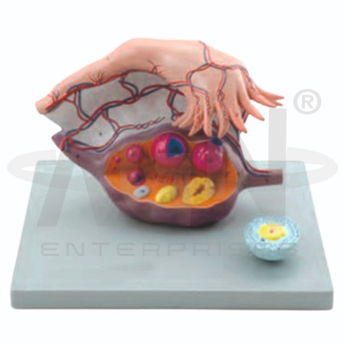
The Ovary Structure Model from AGN Enterprises is a three-dimensional anatomical teaching model designed to demonstrate the internal structure of the human ovary and different stages of ovarian follicle development. The model provides students with a clear visual representation of ovarian anatomy, helping them understand follicular maturation, ovulation, corpus luteum formation, and the reproductive role of the ovaries. It is suitable for medical colleges, nursing institutes, biology laboratories, paramedical institutions, anatomy departments, and reproductive-health education.
The Ovary Structure Model provides an enlarged representation of the ovary, making internal structures and developmental stages easier to identify.
Depending on the specific model configuration, it may demonstrate:
Ovarian Cortex
Ovarian Medulla
Developing Follicles
Mature Follicle
Ovulation
Corpus Luteum
Corpus Albicans
Blood Vessels
The model helps learners visualize how follicles develop within the ovary and how ovarian structures change during the reproductive cycle.
The ovaries are paired organs of the female reproductive system. They contain ovarian follicles at different developmental stages and have important reproductive and endocrine functions.
The ovary can broadly be studied in terms of:
Cortex – the outer region where ovarian follicles are primarily located.
Medulla – the inner region containing connective tissue, blood vessels, lymphatics, and nerves.
The model allows students to observe the relationship between these regions in a three-dimensional format.
The Ovary Structure Model is particularly useful for demonstrating the progression of ovarian follicles.
Depending on the detail represented, students may study stages progressing from early follicles toward a mature ovarian follicle.
During ovulation, an oocyte is released from the mature follicle. The remaining follicular tissue subsequently develops into the corpus luteum, which has an important hormonal role.
If pregnancy does not occur, the corpus luteum eventually regresses and forms a structure known as the corpus albicans.
The Ovary Structure Model transforms microscopic and internal reproductive anatomy into an enlarged visual learning experience. Students can observe follicular development and relate structural changes in the ovary to reproductive physiology.
Teachers can use the model for lessons involving female reproductive anatomy, ovarian follicles, ovulation, menstrual and ovarian cycles, reproductive physiology, and human development.
The three-dimensional presentation helps improve anatomical identification, visualization, conceptual understanding, and retention.
AGN Enterprises supplies anatomical models, biology teaching aids, and educational laboratory equipment for schools, colleges, medical institutions, nursing programs, and professional training centers.
Our reproductive-system models help teachers demonstrate complex anatomical structures and physiological processes using clear three-dimensional representations.









