
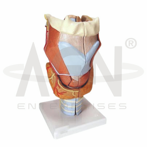
The Larynx Model from AGN Enterprises is a detailed anatomical teaching aid designed to demonstrate the structure of the human larynx and its relationship with the respiratory and vocal systems. The model helps students identify important structures such as the laryngeal cartilages, vocal folds, epiglottis, hyoid region, and upper trachea, depending on the model configuration.
Its three-dimensional design makes complex laryngeal anatomy easier to understand and is suitable for medical colleges, nursing institutes, paramedical institutions, ENT departments, speech and language programs, biology laboratories, and anatomy training centers.
The larynx is located in the neck between the pharynx and trachea. It plays important roles in air passage, voice production, and protection of the lower respiratory tract during swallowing.
Depending on the specific model configuration, structures represented may include:
Epiglottis
Thyroid Cartilage
Cricoid Cartilage
Arytenoid Cartilages
Vocal Folds
Vestibular Folds
Hyoid Bone
Upper Trachea
Associated Muscles and Ligaments
The exact anatomical details may vary according to the supplied model.
The framework of the larynx consists primarily of cartilages connected by membranes, ligaments, and muscles.
The thyroid cartilage forms a major part of the anterior and lateral laryngeal framework, while the cricoid cartilage forms a complete ring below it.
The arytenoid cartilages are particularly important because of their relationship with the vocal folds and their role in controlling the opening and closing of the glottis.
The Larynx Model is useful for demonstrating the position of the vocal folds within the laryngeal cavity.
During phonation, airflow from the lungs causes the appropriately positioned vocal folds to vibrate, producing sound. Movements of the laryngeal muscles alter the position and tension of these folds and contribute to changes in voice production.
This makes the teaching aid valuable for anatomy as well as introductory speech and voice education.
The epiglottis is an elastic cartilage located near the entrance to the larynx.
During swallowing, coordinated movements of the laryngeal structures help protect the airway and direct food and liquids toward the esophagus rather than the lower respiratory tract.
The three-dimensional representation helps students understand the close relationship between the larynx, pharynx, and upper airway.
Inferiorly, the larynx continues into the trachea, forming an important part of the respiratory passage.
Students can use this anatomical teaching aid to trace the general airway pathway:
Pharynx → Larynx → Trachea → Bronchi → Lungs
This helps connect regional laryngeal anatomy with the broader respiratory system.
The human larynx performs several important functions:
Respiration – maintains an airway for the passage of air.
Phonation – contributes to sound and voice production.
Airway Protection – helps prevent material from entering the lower respiratory tract during swallowing.
These functions make laryngeal anatomy particularly important in ENT, respiratory medicine, speech therapy, and clinical anatomy.
This anatomical teaching aid provides students with a three-dimensional approach to studying structures that can be difficult to visualize using diagrams alone.
Teachers can use it for lessons involving laryngeal anatomy, respiratory anatomy, vocal folds, phonation, airway protection, ENT anatomy, and speech physiology.
The model supports anatomical identification, spatial visualization, practical demonstrations, and revision.
AGN Enterprises supplies anatomical models, pathological models, biological teaching aids, and educational laboratory equipment for medical colleges, nursing institutes, paramedical institutions, schools, and professional training centers.
Our anatomical teaching models provide clear visual representations that support practical and classroom-based human-anatomy education.









