
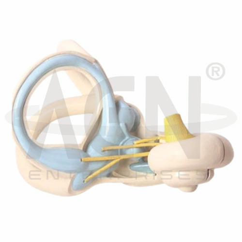
The Labyrinth Model from AGN Enterprises is a detailed anatomical teaching aid designed to demonstrate the internal structures of the human inner ear responsible for hearing and balance. The enlarged three-dimensional model provides a clear representation of the semicircular canals, vestibular region, cochlea, and associated nerve structures.
The model helps students understand the complex spatial arrangement of the vestibular and auditory apparatus, which can be difficult to visualize using two-dimensional diagrams.
It is suitable for medical colleges, nursing institutes, ENT departments, audiology programs, anatomy laboratories, paramedical institutions, biology departments, and health-science training centers.
The Labyrinth Model provides an enlarged three-dimensional representation of important structures of the inner ear.
Depending on the specific model configuration, represented structures may include:
Semicircular Canals
Vestibule
Cochlea
Vestibular Structures
Auditory Structures
Vestibular Nerve
Cochlear Nerve
Vestibulocochlear Nerve Components
Associated Inner Ear Structures
The exact structures, colors, magnification, removable components, and anatomical details may vary according to the supplied model.
The labyrinth is a complex system located within the inner ear.
It contains structures associated with two major sensory functions:
Hearing
Balance and Spatial Orientation
The model provides an enlarged view that allows learners to examine the relationship between the cochlea, vestibule, and semicircular canals.
The semicircular canals form a characteristic looped arrangement within the vestibular apparatus.
There are three semicircular canals oriented in different planes:
Anterior Semicircular Canal
Posterior Semicircular Canal
Lateral Semicircular Canal
Their arrangement enables the vestibular system to detect rotational movements of the head.
The vestibule forms the central region of the inner ear between the cochlea and semicircular canals.
The vestibular apparatus includes sensory structures involved in detecting head position and linear acceleration.
The model helps students understand the central position of this region within the labyrinth.
The cochlea is the spiral-shaped component of the inner ear associated with hearing.
Sound-induced mechanical vibrations are ultimately converted into neural signals by specialized sensory structures within the cochlea.
The distinctive spiral configuration makes the cochlea one of the most recognizable structures on the Labyrinth Model.
The vestibular apparatus contributes to the body’s ability to maintain balance, posture, and spatial orientation.
It includes structures associated with the semicircular canals and vestibular region.
The model provides a useful three-dimensional reference for understanding how these components are positioned relative to one another.
The vestibulocochlear nerve (cranial nerve VIII) carries sensory information from the inner ear toward the brain.
It has two major functional components:
Vestibular Component – associated with balance
Cochlear Component – associated with hearing
Where represented, the nerve structures help students connect inner-ear anatomy with the central nervous system.
The cochlear portion of the labyrinth plays a central role in auditory perception.
A simplified pathway can be described as:
Sound Vibrations → Inner Ear → Cochlear Sensory Structures → Cochlear Nerve → Brain
The model provides the anatomical foundation needed to explain this sensory pathway.
The vestibular portion of the inner ear detects changes in head position and movement.
The semicircular canals are particularly important for detecting angular acceleration, while vestibular structures associated with the utricle and saccule contribute to sensing linear acceleration and head position relative to gravity.
This makes the Labyrinth Model useful for explaining the anatomical basis of equilibrium.
One of the principal educational advantages of the model is its enlarged three-dimensional design.
The semicircular canals occupy different planes, while the cochlea and vestibular structures have complex relationships that can be difficult to appreciate from textbook illustrations.
The model allows these structures to be examined from multiple viewing angles.
The Labyrinth Model can provide an anatomical reference for introductory lessons involving hearing loss, vertigo, vestibular dysfunction, balance disorders, cochlear function, and inner-ear anatomy.
It is particularly useful for ENT, audiology, neurology, and sensory-system education.
The model is intended for anatomical education rather than diagnosis.
The Labyrinth Model provides students with an enlarged three-dimensional approach to studying inner-ear anatomy.
Teachers can use it for lessons involving semicircular canals, cochlea, vestibule, vestibular apparatus, vestibulocochlear nerve, hearing, balance, and equilibrium.
It supports anatomical identification, spatial visualization, practical demonstrations, and examination preparation.
AGN Enterprises supplies ear models, sensory-organ models, neurological teaching aids, anatomical models, scientific instruments, and educational laboratory equipment for medical colleges, nursing institutes, audiology programs, universities, and professional training centers.
Our anatomical teaching models provide clear three-dimensional representations for practical human anatomy and sensory-system education.









