
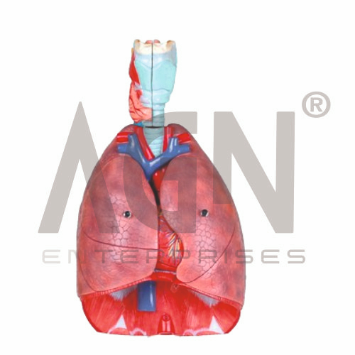
The Larynx, Heart and Lung Model from AGN Enterprises is a comprehensive anatomical teaching aid designed to demonstrate the major structures of the human respiratory system together with the heart and associated thoracic anatomy. By presenting the larynx, trachea, lungs, and heart together, the model helps students understand the anatomical relationship between the respiratory and cardiovascular systems.
The three-dimensional presentation is suitable for medical colleges, nursing institutes, paramedical institutions, schools, biology laboratories, anatomy departments, respiratory therapy programs, and health-science training centers.
This teaching model provides an integrated view of important organs involved in breathing, gas exchange, circulation, and voice production.
Depending on the specific model configuration, structures represented may include:
Larynx
Trachea
Right and Left Main Bronchi
Right Lung
Left Lung
Heart
Major Blood Vessels
Pulmonary Vessels
Associated Thoracic Structures
The exact anatomical details and removable components may vary according to the supplied model.
The larynx is located between the pharynx and trachea and plays important roles in respiration, voice production, and airway protection.
Where sufficient detail is represented, students may identify structures such as the epiglottis, thyroid cartilage, cricoid cartilage, and vocal region.
Including the larynx with the lungs helps learners trace the respiratory pathway from the upper airway toward the thorax.
The trachea carries air inferiorly from the larynx before dividing into the right and left main bronchi.
Students can use the Larynx, Heart and Lung Model to understand the general pathway:
Larynx → Trachea → Main Bronchi → Lungs
This provides a clear visual foundation for studying the organization of the human respiratory tract.
The lungs are the principal organs of respiration and contain the structures where gas exchange ultimately occurs.
The right lung normally has three lobes, while the left lung normally has two lobes and accommodates the position of the heart.
Where lobar divisions are represented, the model helps students compare the external anatomy of the right and left lungs.
The heart is positioned within the thoracic cavity between the lungs, primarily in the middle mediastinum.
The model allows learners to observe the spatial relationship between the heart and lungs. Depending on its construction, major external cardiac structures or vessels may also be represented.
This integrated presentation is particularly useful when introducing cardiopulmonary anatomy.
The respiratory and cardiovascular systems work closely together to support gas exchange and transport gases throughout the body.
Blood is pumped from the right side of the heart toward the lungs through the pulmonary circulation. After gas exchange, oxygenated blood returns to the left side of the heart and is subsequently pumped into the systemic circulation.
Studying the heart and lungs together helps students connect anatomical organization with basic physiology.
Teachers can use the model to explain the general movement of inhaled air through the respiratory tract:
Larynx → Trachea → Bronchi → Smaller Airways → Lungs
This pathway helps learners understand how structures of the upper and lower respiratory systems form a continuous conducting system.
This anatomical teaching aid combines several major thoracic and respiratory structures into one visual model. Students can study the position of the larynx, trachea, bronchi, lungs, and heart while understanding how these organs relate to one another.
It is useful for lessons involving human anatomy, respiratory anatomy, cardiovascular anatomy, cardiopulmonary anatomy, pulmonary circulation, airway anatomy, and basic physiology.
The model supports anatomical identification, spatial understanding, practical demonstrations, and classroom revision.
AGN Enterprises supplies anatomical models, pathological models, biological teaching aids, and educational laboratory equipment for medical colleges, nursing institutes, paramedical institutions, schools, and professional training centers.
Our anatomical teaching aids help learners understand complex human-body systems through clear three-dimensional representations.









