
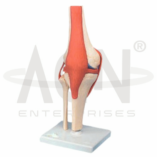
The Knee Joints Set from AGN Enterprises is a detailed anatomical teaching aid designed to demonstrate the structure, movement, and important components of the human knee joint. The set helps students study the relationship between the femur, tibia, patella, menisci, ligaments, and other structures involved in knee stability and movement.
It is suitable for medical colleges, nursing institutes, physiotherapy colleges, orthopedic departments, paramedical institutions, sports-science programs, anatomy laboratories, and health-science training centers.
The knee is a complex synovial joint that primarily connects the femur and tibia, with the patella forming the patellofemoral articulation.
Depending on the specific configuration, structures represented in the set may include:
Femur
Tibia
Fibula
Patella
Medial Meniscus
Lateral Meniscus
Anterior Cruciate Ligament (ACL)
Posterior Cruciate Ligament (PCL)
Collateral Ligaments
Associated Joint Structures
The exact number of models, structures, and anatomical details may vary according to the supplied set.
The major bones associated with the knee region include the femur, tibia, fibula, and patella.
The distal femur articulates with the proximal tibia, while the patella articulates with the femur anteriorly.
The three-dimensional model helps students understand how these bones are positioned relative to one another.
The anterior cruciate ligament (ACL) and posterior cruciate ligament (PCL) are important stabilizing structures within the knee.
The ACL helps resist excessive anterior translation of the tibia relative to the femur, while the PCL helps resist excessive posterior translation.
Where represented, the model allows students to examine their orientation and anatomical relationship within the joint.
The collateral ligaments contribute to stability on the medial and lateral sides of the knee.
These include the:
Medial Collateral Ligament (MCL)
Lateral Collateral Ligament (LCL)
The model provides a useful visual reference for understanding their positions and roles in stabilizing the knee.
The knee contains medial and lateral menisci, which are fibrocartilaginous structures located between the femoral condyles and tibial plateau.
They contribute to load distribution, joint congruence, shock absorption, and stability.
Where represented, students can compare the shape and position of the two menisci.
The knee primarily permits flexion and extension, with limited rotational movement possible in certain positions.
The Knee Joints Set can help teachers explain how the bones, ligaments, and other structures work together during joint movement.
This makes the set particularly useful for physiotherapy, orthopedics, biomechanics, and sports-science education.
The Knee Joints Set provides students with a three-dimensional approach to studying one of the body’s most important weight-bearing joints.
Teachers can use it for lessons involving knee anatomy, joint mechanics, ligaments, menisci, musculoskeletal anatomy, biomechanics, physiotherapy, and orthopedics.
The set supports anatomical identification, spatial visualization, practical demonstrations, and examination preparation.
AGN Enterprises supplies anatomical models, joint models, skeletal teaching aids, biological models, and educational laboratory equipment for medical colleges, nursing institutes, physiotherapy programs, paramedical institutions, and professional training centers.
Our anatomical teaching aids provide clear three-dimensional representations for practical and classroom-based anatomy education.









