
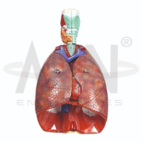
The Human Respiratory System Model from AGN Enterprises is a detailed anatomical teaching aid designed to demonstrate the major organs and structures involved in human respiration. The model provides a three-dimensional representation of the lungs, trachea, bronchial structures, laryngeal region, and diaphragm, helping students understand the anatomy and organization of the respiratory tract.
The model is suitable for medical colleges, nursing institutes, physiotherapy colleges, paramedical institutions, biology departments, anatomy laboratories, respiratory-care education, and health-science training centers.
The Human Respiratory System Model provides a clear representation of important structures associated with breathing and pulmonary anatomy.
Depending on the specific model configuration, represented structures may include:
Laryngeal Region
Trachea
Right Lung
Left Lung
Main Bronchi
Bronchial Structures
Pulmonary Blood Vessels
Diaphragm
Associated Respiratory Structures
The exact structures, removable components, colors, dimensions, and anatomical details may vary according to the supplied model.
The respiratory system is responsible for ventilation and gas exchange, supplying the body with oxygen while helping eliminate carbon dioxide.
Air travels through a series of conducting passages before reaching the gas-exchange regions of the lungs.
The model helps students connect individual respiratory organs with the overall process of breathing.
The larynx forms an important part of the upper respiratory tract and connects the pharyngeal region with the trachea.
It participates in air conduction, phonation, and protection of the lower airway.
The model helps learners understand its position above the trachea and lungs.
The trachea, commonly called the windpipe, conducts air toward the lungs.
Its wall is supported by cartilaginous structures that help maintain an open airway.
Inferiorly, the trachea divides into the right and left main bronchi.
The right and left main bronchi carry air from the trachea into the corresponding lungs.
Within the lungs, the bronchi progressively branch into smaller airways, ultimately leading toward the respiratory regions where gas exchange occurs.
The model provides a useful three-dimensional reference for studying this branching arrangement.
The right lung is normally divided into three lobes:
Superior Lobe
Middle Lobe
Inferior Lobe
These lobes are separated by horizontal and oblique fissures.
The model helps students distinguish the right lung from the left and understand their anatomical differences.
The left lung is normally divided into two lobes:
Superior Lobe
Inferior Lobe
The left lung accommodates the position of the heart within the thoracic cavity and therefore differs in shape and organization from the right lung.
The model provides a clear visual comparison of both lungs.
The lungs have an extensive vascular network associated with pulmonary circulation.
The pulmonary arteries carry deoxygenated blood from the right side of the heart toward the lungs, while the pulmonary veins return oxygenated blood to the left side of the heart.
Where represented, these vessels help connect respiratory anatomy with cardiovascular physiology.
The diaphragm is the principal muscle of respiration and forms a muscular partition between the thoracic and abdominal cavities.
During inspiration, contraction of the diaphragm increases the volume of the thoracic cavity and contributes to drawing air into the lungs.
The diaphragm is clearly represented at the inferior aspect of the model.
The model can be used to explain the basic mechanics of respiration.
During inspiration, contraction of the diaphragm and other respiratory muscles increases thoracic volume, allowing air to enter the lungs.
During expiration, changes in respiratory muscle activity and elastic recoil contribute to movement of air out of the lungs.
A simplified pathway of inspired air can be demonstrated as:
Upper Airway → Larynx → Trachea → Main Bronchi → Smaller Bronchi → Bronchioles → Alveolar Regions
This pathway helps students connect the visible anatomical structures with the physiological process of ventilation.
Gas exchange occurs primarily across the thin respiratory surfaces of the alveoli and surrounding pulmonary capillaries.
Oxygen moves from inhaled air into the blood, while carbon dioxide moves from the blood toward the alveolar air for elimination during expiration.
Although microscopic alveolar anatomy may not be represented on this model, the model provides the structural context necessary for understanding pulmonary gas exchange.
The Human Respiratory System Model provides an anatomical foundation for introductory lessons involving airway anatomy, pulmonary circulation, lung lobes, respiratory mechanics, bronchial anatomy, and respiratory disorders.
It can be used as a structural reference when discussing conditions affecting the lungs and airways.
The model is intended for educational demonstration rather than diagnosis.
The Human Respiratory System Model provides students with a three-dimensional approach to studying respiratory anatomy.
Teachers can use it for lessons involving lungs, trachea, bronchi, larynx, pulmonary vessels, diaphragm, breathing mechanics, and gas exchange.
It supports anatomical identification, spatial visualization, practical demonstrations, and examination preparation.
AGN Enterprises supplies respiratory-system models, lung models, anatomical teaching aids, biological models, scientific instruments, and educational laboratory equipment for medical colleges, nursing institutes, physiotherapy programs, universities, and professional training centers.
Our anatomical teaching models provide clear three-dimensional representations for practical human anatomy and health-science education.









