
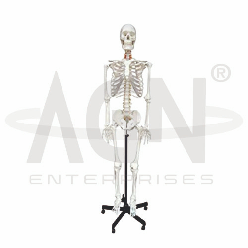
The Human Skeleton Model from AGN Enterprises is a detailed anatomical teaching aid designed to demonstrate the bones, major joints, and overall organization of the human skeletal system. It provides students with a three-dimensional representation for studying bone identification, skeletal relationships, body support, and joint anatomy.
The model is suitable for medical colleges, nursing institutes, physiotherapy colleges, paramedical institutions, schools, biology laboratories, anatomy departments, orthopedic education, and health-science training centers.
The adult human skeleton typically consists of 206 bones, organized into the axial and appendicular skeletons.
Depending on the specific model configuration, structures represented may include:
Skull
Vertebral Column
Rib Cage
Sternum
Shoulder Girdle
Upper Limbs
Pelvis
Lower Limbs
Major Joints
Hands and Feet
The exact construction, articulation, mounting system, and movable components may vary according to the supplied model.
The axial skeleton forms the central framework of the body and includes the skull, vertebral column, ribs, and sternum.
These structures provide support and protection for important organs and tissues, including the brain, spinal cord, heart, and lungs.
The model helps students identify individual axial bones and understand their anatomical arrangement.
The appendicular skeleton includes the bones of the upper and lower limbs together with the pectoral and pelvic girdles.
It plays an important role in movement and interaction with the surrounding environment.
The three-dimensional skeleton allows students to study the relationship between limb bones, girdles, and major joints.
The skull forms the skeletal framework of the head and protects the brain.
It consists of cranial and facial bones that contribute to structures such as the cranial cavity, orbits, nasal cavity, and jaws.
Where movable or removable components are provided, students can examine selected skull structures in greater detail.
The vertebral column forms the central supporting axis of the trunk.
It includes the:
Cervical Vertebrae
Thoracic Vertebrae
Lumbar Vertebrae
Sacrum
Coccyx
The model helps learners understand spinal regions, natural curvatures, and the relationship of the vertebral column to the skull, ribs, and pelvis.
The upper limbs include the humerus, radius, ulna, wrist bones, metacarpals, and phalanges.
The lower limbs include the femur, patella, tibia, fibula, ankle bones, metatarsals, and phalanges.
Studying these structures on a complete skeleton helps students understand limb organization and major joint relationships.
Depending on the construction, the Human Skeleton Model may allow demonstration of movement at major articulated joints.
Important regions include the:
Shoulder
Elbow
Wrist
Hip
Knee
Ankle
This makes the model particularly useful for anatomy, physiotherapy, orthopedics, and biomechanics education.
The skeletal system performs several essential functions, including:
Body Support
Protection of Internal Organs
Movement with Skeletal Muscles
Mineral Storage
Blood Cell Formation in Bone Marrow
Providing Structural Shape to the Body
The model helps students connect skeletal anatomy with these important physiological functions.
The Human Skeleton Model provides a comprehensive three-dimensional approach to studying human osteology.
Teachers can use it for lessons involving skeletal anatomy, bone identification, vertebral anatomy, joint anatomy, musculoskeletal anatomy, posture, movement, physiotherapy, and orthopedics.
It supports anatomical identification, practical demonstrations, spatial visualization, and examination preparation.
AGN Enterprises supplies anatomical models, skeleton models, joint models, biological teaching aids, and educational laboratory equipment for medical colleges, nursing institutes, physiotherapy programs, schools, paramedical institutions, and professional training centers.
Our anatomical models provide clear three-dimensional representations to support practical and classroom-based human anatomy education.









