
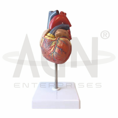
The Human Heart Model from AGN Enterprises is a detailed anatomical teaching aid designed to demonstrate the external and internal anatomy of the human heart, major blood vessels, coronary circulation, chambers, and associated cardiac structures.
The three-dimensional design provides students with a clear understanding of the heart’s structure and its role in the cardiovascular system. The model is mounted on a stable stand for convenient classroom, laboratory, and demonstration use.
It is suitable for medical colleges, nursing institutes, physiotherapy colleges, paramedical institutions, biology departments, anatomy laboratories, cardiology education, and health-science training centers.
The Human Heart Model provides a three-dimensional representation of important structures of the cardiovascular system.
Depending on the specific model configuration, represented structures may include:
Right Atrium
Left Atrium
Right Ventricle
Left Ventricle
Aorta
Pulmonary Trunk
Pulmonary Arteries
Pulmonary Veins
Superior Vena Cava
Inferior Vena Cava
Coronary Arteries
Cardiac Veins
The exact removable components, internal structures, colors, dimensions, and anatomical details may vary according to the supplied model.
The human heart contains four chambers:
Right Atrium
Right Ventricle
Left Atrium
Left Ventricle
The right side primarily receives deoxygenated blood and pumps it toward the lungs, while the left side receives oxygenated blood from the lungs and pumps it into the systemic circulation.
The model helps students understand the positions and relationships of these chambers.
The right atrium receives systemic venous blood primarily through the superior and inferior vena cava.
Blood then passes from the right atrium toward the right ventricle.
Where internal structures are visible or removable, the model provides a useful reference for studying this pathway.
The right ventricle pumps blood into the pulmonary circulation through the pulmonary trunk.
The model helps students visualize its position relative to the left ventricle, atria, and major vessels.
The left atrium receives oxygenated blood returning from the lungs through the pulmonary veins.
Blood then passes into the left ventricle before entering systemic circulation.
The model provides a three-dimensional reference for understanding this important part of the circulatory pathway.
The left ventricle pumps oxygenated blood into the aorta for distribution throughout the body.
Its muscular wall is substantially thicker than that of the right ventricle because it must generate the pressure required for systemic circulation.
Where internal anatomy is shown, this difference can be demonstrated clearly.
The aorta is the largest artery in the human body and carries oxygenated blood from the left ventricle into systemic circulation.
The prominent aortic region visible on the model helps students understand its relationship with the heart and other major vessels.
The right side of the heart pumps blood toward the lungs through the pulmonary trunk and pulmonary arteries.
After gas exchange in the lungs, oxygenated blood returns to the left atrium through the pulmonary veins.
The model can be used to explain this pulmonary circulation pathway in three dimensions.
The coronary arteries supply oxygenated blood to the cardiac muscle itself.
The model visibly demonstrates major coronary vessels running across the external surface of the heart, providing an excellent reference for studying coronary circulation and cardiac vascular anatomy.
Cardiac veins drain blood from the myocardium and contribute to the venous circulation of the heart.
Where represented, they can be distinguished from coronary arteries using anatomical position and conventional color coding.
This helps students study arterial and venous circulation simultaneously.
Where internal structures are represented, the model may demonstrate the major cardiac valves:
Tricuspid Valve
Pulmonary Valve
Mitral Valve
Aortic Valve
These valves help maintain one-way blood flow through the chambers and major vessels of the heart.
The Human Heart Model can be used to demonstrate the basic pathway of blood:
Body → Venae Cavae → Right Atrium → Right Ventricle → Pulmonary Arteries → Lungs → Pulmonary Veins → Left Atrium → Left Ventricle → Aorta → Body
This makes the model especially useful for explaining the relationship between cardiac anatomy and circulation.
The Human Heart Model provides a useful structural foundation for lessons involving cardiac anatomy, coronary circulation, pulmonary circulation, systemic circulation, heart chambers, valves, and major blood vessels.
It can also serve as an anatomical reference when introducing cardiovascular conditions and clinical cardiac terminology.
The model is intended for educational demonstration rather than diagnosis.
The Human Heart Model provides students with a practical three-dimensional approach to studying cardiovascular anatomy.
Teachers can use it for lessons involving heart chambers, coronary arteries, cardiac veins, aorta, pulmonary vessels, cardiac valves, and blood circulation.
It supports anatomical identification, spatial visualization, practical demonstrations, and examination preparation.
AGN Enterprises supplies heart models, cardiovascular models, anatomical teaching aids, biological models, scientific instruments, and educational laboratory equipment for medical colleges, nursing institutes, universities, hospitals, and professional training centers.
Our anatomical teaching models provide clear three-dimensional representations for practical human anatomy and cardiovascular education.









