
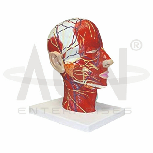
The Human Head Model from AGN Enterprises is a detailed anatomical teaching aid designed to demonstrate the muscles, arteries, veins, nerves, and other important structures of the human head and upper neck. The color-coded three-dimensional design helps students identify complex anatomical relationships more easily than with conventional diagrams.
The model is particularly useful for studying facial musculature, superficial blood vessels, cranial nerve branches, scalp anatomy, and cervical structures.
It is suitable for medical colleges, nursing institutes, physiotherapy colleges, dental colleges, anatomy departments, paramedical institutions, universities, and health-science training centers.
The Human Head Model provides a three-dimensional representation of important superficial structures of the head and neck.
Depending on the specific model configuration, represented structures may include:
Facial Muscles
Muscles of Mastication
Scalp Muscles
Facial Arteries
Superficial Arteries
Veins
Facial Nerves
Sensory Nerve Branches
External Ear
Eye and Orbital Region
Nasal Region
Oral Region
Upper Neck Muscles
The exact structures, labeling, colors, dimensions, and anatomical details may vary according to the supplied model.
The model demonstrates important muscles associated with facial expression.
These muscles contribute to movements of the eyebrows, eyelids, nose, lips, cheeks, and mouth and play an essential role in facial expression and communication.
The three-dimensional presentation helps learners understand their locations and relationships with superficial nerves and blood vessels.
Muscles associated with mastication produce movements of the mandible required for chewing.
Where represented, the model can help students study important muscles such as the masseter and temporalis and understand their relationship with the skull, jaw, and surrounding facial structures.
The head receives an extensive arterial blood supply.
The model uses clearly differentiated vascular structures to help demonstrate important superficial arterial pathways of the face, scalp, and temporal regions.
Where represented, students can trace major vessels and observe their relationships with facial muscles and nerves.
The superficial veins of the head and face form an extensive drainage network.
Where represented, these vessels provide students with a useful three-dimensional reference for studying venous anatomy alongside the corresponding arteries.
Color differentiation helps distinguish veins from arteries and surrounding anatomical structures.
Numerous nerve branches are distributed throughout the face and scalp.
Where represented, the model helps students understand the pathways and distribution of important motor and sensory nerves of the head.
This makes it useful for neuroanatomy, dental anatomy, and clinical head-and-neck education.
The facial nerve (cranial nerve VII) provides motor innervation to the muscles of facial expression and has additional specialized functions.
Its terminal branches spread across different regions of the face.
Where displayed, the model provides a useful visual reference for studying the distribution of these branches relative to facial muscles.
The trigeminal nerve (cranial nerve V) is the major sensory nerve of the face and also provides motor innervation to muscles involved in mastication.
Its three principal divisions are:
Ophthalmic Division (V1)
Maxillary Division (V2)
Mandibular Division (V3)
Where represented, the model can support teaching of their general distribution across the facial region.
The model demonstrates the external anatomy of the head and can be used to explain relationships between the scalp, muscles, blood vessels, nerves, skull, and facial structures.
This is especially useful for regional anatomy lessons in which students need to understand structures from a lateral or superficial perspective.
The model provides anatomical landmarks around the eye, nose, ear, and mouth.
These regions help students orient themselves when studying surrounding muscles, vessels, and nerves.
The visible external structures also make the model suitable for introductory clinical and regional anatomy demonstrations.
The head is anatomically continuous with the neck through a complex arrangement of muscles, vessels, nerves, skeletal structures, and visceral pathways.
The model includes the upper cervical region, helping learners understand how structures of the face and scalp continue toward the neck.
The structures represented on the Human Head Model contribute to numerous functions, including:
The Human Head Model provides a useful anatomical reference for introductory teaching involving facial nerve pathways, trigeminal nerve distribution, facial muscles, vascular anatomy, dental anatomy, and head-and-neck examination.
It can help learners understand the spatial relationships between muscles, nerves, and blood vessels before progressing to more advanced clinical anatomy.
The model is intended for educational demonstration rather than diagnosis.
The Human Head Model provides students with a clear three-dimensional approach to studying complex head anatomy.
Teachers can use it for lessons involving facial muscles, arteries, veins, nerves, scalp structures, mastication, facial expression, and regional head-and-neck anatomy.
The color-coded design supports anatomical identification, spatial visualization, practical demonstrations, and examination preparation.
AGN Enterprises supplies head and neck models, muscular models, vascular models, neurological teaching aids, anatomical models, scientific instruments, and educational laboratory equipment for medical colleges, nursing institutes, dental colleges, physiotherapy programs, universities, and professional training centers.
Our anatomical teaching models provide clear three-dimensional representations for practical human anatomy and medical education.









