
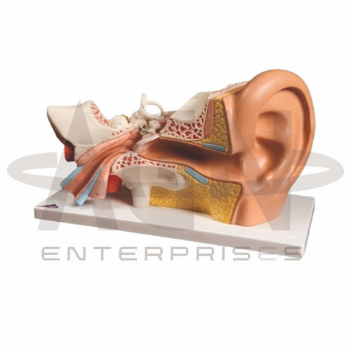
The Human Ear Model from AGN Enterprises is a detailed anatomical teaching aid designed to demonstrate the external, middle, and inner structures of the human ear. The model provides a three-dimensional representation of important components involved in hearing and balance, helping students understand their anatomical positions and functional relationships.
It is suitable for medical colleges, nursing institutes, audiology programs, ENT departments, paramedical institutions, schools, biology laboratories, anatomy departments, and health-science training centers.
The Human Ear Model provides an enlarged representation of the major anatomical regions of the ear.
Depending on the specific model configuration, represented structures may include:
Auricle (Pinna)
External Auditory Canal
Tympanic Membrane
Malleus
Incus
Stapes
Eustachian Tube
Cochlea
Semicircular Canals
Vestibule
Auditory Nerve
The exact structures, removable components, colors, magnification, and anatomical details may vary according to the supplied model.
The external ear consists primarily of the auricle and external auditory canal.
The auricle collects sound waves and directs them through the auditory canal toward the tympanic membrane.
The model helps students understand the external pathway through which sound enters the auditory system.
The tympanic membrane, commonly known as the eardrum, separates the external ear from the middle ear.
Sound waves cause the tympanic membrane to vibrate, initiating mechanical transmission through the auditory ossicles.
Where represented, the model clearly demonstrates its location within the ear.
The middle ear is an air-filled cavity containing three small auditory ossicles:
Malleus
Incus
Stapes
These bones transmit vibrations from the tympanic membrane toward the inner ear.
The enlarged model helps learners identify each ossicle and understand their arrangement.
The Eustachian tube, also called the auditory or pharyngotympanic tube, connects the middle-ear cavity with the nasopharyngeal region.
It contributes to pressure equalization across the tympanic membrane.
Where represented, the model helps students visualize its orientation relative to the middle ear.
The inner ear contains structures involved in hearing and equilibrium.
Important components include the:
Cochlea
Vestibule
Semicircular Canals
These structures form part of a complex sensory system that converts mechanical stimuli into neural information.
The cochlea is a spiral-shaped structure associated with hearing.
It contains specialized sensory structures responsible for converting sound-related mechanical vibrations into electrical signals that can be transmitted through neural pathways.
The model provides a clear three-dimensional representation of its characteristic shape and position.
The semicircular canals form part of the vestibular apparatus and contribute to the detection of rotational movements of the head.
Their distinctive three-dimensional arrangement makes them particularly suitable for study using an anatomical model.
Students can examine their relationship with the cochlea and other inner-ear structures.
The human ear performs two major sensory functions:
The model helps students connect anatomical structures with these important sensory functions.
The Human Ear Model provides students with a three-dimensional approach to studying auditory and vestibular anatomy.
Teachers can use it for lessons involving external ear anatomy, middle ear, auditory ossicles, cochlea, semicircular canals, hearing, balance, audiology, and ENT anatomy.
The model supports anatomical identification, spatial visualization, practical demonstrations, and examination preparation.
AGN Enterprises supplies anatomical models, sensory-organ models, ear models, biological teaching aids, scientific instruments, and educational laboratory equipment for medical colleges, nursing institutes, schools, universities, and professional training centers.
Our anatomical teaching models provide clear three-dimensional representations for practical and classroom-based human anatomy education.









