
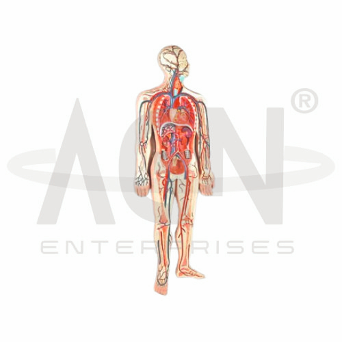
The Human Circulatory System Model from AGN Enterprises is a detailed anatomical teaching aid designed to demonstrate the heart, major arteries, veins, and pathways of blood circulation throughout the human body. It helps students understand how the cardiovascular system transports oxygen, nutrients, hormones, and other substances while carrying metabolic waste products away from tissues.
The model is suitable for medical colleges, nursing institutes, paramedical institutions, schools, biology laboratories, anatomy departments, physiology laboratories, and health-science training centers.
The circulatory system consists primarily of the heart and an extensive network of blood vessels.
Depending on the specific model configuration, structures represented may include:
Heart
Aorta
Major Arteries
Major Veins
Venae Cavae
Pulmonary Arteries
Pulmonary Veins
Systemic Circulation
Pulmonary Circulation
The exact vessels, anatomical details, colors, and structures represented may vary according to the supplied model.
The heart is the muscular pumping organ at the center of the cardiovascular system.
It consists of four chambers:
Right Atrium
Right Ventricle
Left Atrium
Left Ventricle
The right side primarily pumps blood toward the lungs, while the left side pumps oxygenated blood into the systemic circulation.
Arteries generally carry blood away from the heart.
The aorta is the principal artery of systemic circulation and gives rise to branches that distribute oxygenated blood throughout the body.
The model helps students trace major arterial pathways and understand how blood reaches different organs and body regions.
Veins generally return blood toward the heart.
Major systemic veins ultimately drain into the superior and inferior vena cava, which return deoxygenated blood to the right atrium.
The model provides a useful visual comparison between major arterial and venous pathways.
Pulmonary circulation transports blood between the heart and lungs.
A simplified pathway is:
Right Ventricle → Pulmonary Arteries → Lungs → Pulmonary Veins → Left Atrium
In the lungs, blood participates in gas exchange before returning to the heart.
Systemic circulation distributes oxygenated blood from the left side of the heart to tissues throughout the body and returns deoxygenated blood to the right side.
A simplified pathway is:
Left Ventricle → Aorta → Arteries → Body Tissues → Veins → Venae Cavae → Right Atrium
The model helps students understand this continuous circulation in a three-dimensional anatomical context.
The Human Circulatory System Model can be used to demonstrate the overall direction of blood flow:
Body → Right Heart → Lungs → Left Heart → Body
This provides students with a clear foundation for understanding the relationship between pulmonary and systemic circulation.
The circulatory system performs several essential functions, including:
Transporting Oxygen and Nutrients
Removing Carbon Dioxide and Metabolic Wastes
Transporting Hormones
Supporting Temperature Regulation
Contributing to Immune Defense
Maintaining Internal Physiological Balance
The model helps connect these physiological functions with cardiovascular anatomy.
The Human Circulatory System Model provides students with a visual approach to understanding the extensive network of blood vessels throughout the body.
Teachers can use it for lessons involving heart anatomy, arteries, veins, blood flow, pulmonary circulation, systemic circulation, cardiovascular physiology, and human anatomy.
The model supports anatomical identification, circulation tracing, practical demonstrations, and examination preparation.
AGN Enterprises supplies anatomical models, biological teaching aids, cardiovascular models, pathological models, scientific instruments, and educational laboratory equipment for schools, medical colleges, nursing institutes, paramedical institutions, and training centers.
Our anatomical models provide clear three-dimensional representations to support practical and classroom-based human anatomy education.









