
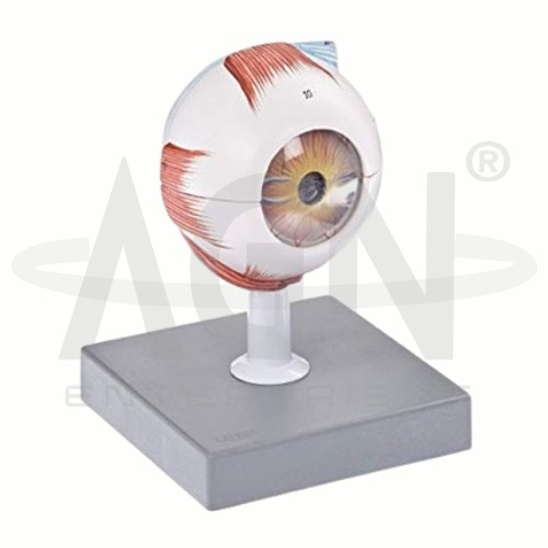
This 6X life-size eyeball model is designed to provide a detailed study of ocular anatomy with detachable parts for closer examination. It clearly demonstrates the three main layers of the eye: the tunica externa, showing the cornea and sclera with attachments of ocular muscles and the optic nerve; the tunica media, including the iris, ciliary body, and choroid; and the tunica interna, representing the retina. The model also highlights the refractive media of the eye, including the lens and vitreous body. Mounted on a board, it serves as an effective educational tool for understanding the structure and function of the human eye.









