
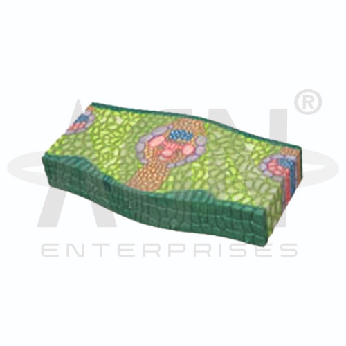
The Dicot Herbaceous Stem Section (Helianthus Annuus) Model from AGN Enterprises is an enlarged botanical teaching aid designed to demonstrate the internal anatomy and tissue arrangement of a typical dicot herbaceous stem using sunflower (Helianthus annuus) as an example.
The three-dimensional model helps students identify the epidermis, cortex, vascular bundles, xylem, phloem, cambium, medullary rays, and pith while understanding their relative positions within the stem.
It is suitable for schools, colleges, universities, botany laboratories, biology departments, agricultural institutes, horticulture programs, and plant-science training centers.
The Dicot Herbaceous Stem Section (Helianthus Annuus) Model provides an enlarged representation of the tissues visible in a transverse section of a young sunflower stem.
Depending on the specific model configuration, represented structures may include:
Epidermis
Hypodermis
Cortex
Endodermis
Pericycle
Phloem
Vascular Cambium
Xylem
Medullary Rays
Pith
The exact structures, colors, magnification, and sectional details may vary according to the supplied model.
The epidermis forms the outer protective layer of the young stem.
It helps protect the underlying tissues from mechanical injury and excessive water loss. A cuticle may cover the epidermal surface.
The enlarged model makes this outermost tissue layer easy to distinguish from the internal cortical tissues.
Beneath the epidermis lies the cortical region.
In a typical young dicot stem such as Helianthus, the hypodermal region commonly contains collenchymatous tissue, which contributes to mechanical support while allowing flexibility.
Deeper cortical tissues perform functions associated with storage and other physiological processes.
The innermost region of the cortex is associated with the endodermis, which separates cortical tissues from the vascular region.
Internal to this lies the pericycle region.
Where represented, the model helps students identify these tissues and understand their positions relative to the vascular bundles.
A characteristic feature of a dicot stem is the arrangement of vascular bundles in a ring around the central pith.
In a typical young Helianthus stem, the vascular bundles are conjoint, collateral, and open, with cambium present between the phloem and xylem.
This arrangement is an important feature for distinguishing dicot stems from many monocot stems.
The phloem is positioned toward the outer portion of each vascular bundle.
It transports organic substances, particularly sugars produced through photosynthesis, to different regions of the plant.
The model helps learners identify the phloem and compare its position with the xylem.
The vascular cambium lies between the phloem and xylem in an open vascular bundle.
Cambial activity can produce secondary xylem toward the inside and secondary phloem toward the outside, contributing to secondary growth.
The model provides a useful introduction to the developmental processes responsible for increasing stem thickness.
The xylem occupies the inner portion of each vascular bundle.
Its principal functions include transporting water and dissolved mineral nutrients and providing structural support.
The model helps students recognize the orientation of xylem relative to the cambium, phloem, and central pith.
Medullary rays occur between vascular bundles and extend radially between the central and peripheral regions of the stem.
They contribute to radial transport and storage.
Their arrangement is particularly useful when teaching the organization of vascular and ground tissues in a dicot stem.
The pith forms the prominent central region of a typical young sunflower stem.
It consists mainly of parenchymatous tissue and is associated primarily with storage.
The large central pith provides a useful reference point when identifying the ring of surrounding vascular bundles.
Important features commonly demonstrated by the model include:
These characteristics make the model useful for comparing dicot and monocot stem anatomy.
The Dicot Herbaceous Stem Section (Helianthus Annuus) Model provides students with an enlarged three-dimensional approach to studying plant anatomy.
Teachers can use it for lessons involving dicot stem structure, vascular bundles, xylem, phloem, cambium, cortex, pith, medullary rays, secondary growth, and comparative plant anatomy.
The model supports tissue identification, practical demonstrations, laboratory study, and examination preparation.
AGN Enterprises supplies botanical models, plant-anatomy models, biological teaching aids, scientific instruments, and educational laboratory equipment for schools, colleges, universities, agricultural institutes, and professional laboratories.
Our botanical teaching models provide clear three-dimensional representations to support practical botany and biology education.









