
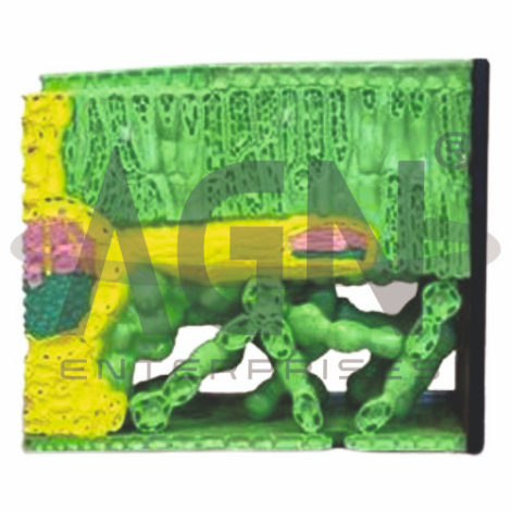
The Dicot Leaf Model from AGN Enterprises is an enlarged botanical teaching aid designed to demonstrate the internal anatomy and tissue organization of a typical dicotyledonous leaf. The model provides a clear three-dimensional representation of the epidermis, mesophyll tissues, vascular bundle, stomata, and other structures involved in photosynthesis, gas exchange, and transport.
It is suitable for schools, colleges, universities, botany laboratories, biology departments, agricultural institutes, horticulture programs, and plant-science training centers.
The Dicot Leaf Model demonstrates the characteristic internal organization of a typical dorsiventral dicot leaf.
Depending on the specific model configuration, represented structures may include:
Cuticle
Upper Epidermis
Palisade Mesophyll
Spongy Mesophyll
Intercellular Air Spaces
Vascular Bundle
Xylem
Phloem
Lower Epidermis
Stomata and Guard Cells
The exact structures, colors, magnification, and sectional details may vary according to the supplied model.
The outer surface of the leaf is generally covered by a protective cuticle.
Immediately beneath it lies the upper epidermis, which forms a protective cellular layer and allows light to reach the photosynthetic tissues below.
The enlarged model helps students distinguish these superficial structures from the underlying mesophyll.
The palisade mesophyll is located beneath the upper epidermis in a typical dorsiventral dicot leaf.
Its elongated cells contain numerous chloroplasts and are particularly important for photosynthesis.
The model provides a clear visual representation of the compact arrangement of palisade cells.
The spongy mesophyll is generally positioned below the palisade layer.
Its cells are more loosely arranged, creating numerous intercellular air spaces that facilitate the movement of gases within the leaf.
This structure helps students connect leaf anatomy with photosynthesis and gas exchange.
Leaf veins contain vascular bundles responsible for transport within the plant.
The two principal vascular tissues are:
Xylem – transports water and dissolved mineral nutrients
Phloem – transports sugars and other organic substances
In a typical dicot leaf vascular bundle, xylem is generally oriented toward the upper surface and phloem toward the lower surface.
Stomata are microscopic pores in the epidermis that regulate gas exchange and water loss.
Each stoma is bordered by specialized guard cells, which regulate the opening and closing of the pore.
In many dorsiventral dicot leaves, stomata are more numerous on the lower epidermis.
Where represented, the enlarged model makes these structures easier to identify.
Carbon dioxide enters the leaf through stomata and moves through the internal air spaces toward photosynthetic cells.
Oxygen and water vapor can move outward through the same stomatal openings.
The model therefore provides a useful visual reference for teaching gas exchange, transpiration, and stomatal regulation.
The internal organization of a dicot leaf is closely adapted to photosynthesis.
The palisade mesophyll contains abundant chloroplasts for capturing light, while the spongy mesophyll provides air spaces that facilitate gaseous movement.
The vascular tissues supply water and transport products of photosynthesis to other parts of the plant.
Important anatomical features commonly associated with a typical dorsiventral dicot leaf include:
These features make the model particularly useful for comparison with monocot leaf anatomy.
The Dicot Leaf Model provides an enlarged three-dimensional approach to studying plant anatomy.
Teachers can use it for lessons involving leaf structure, epidermis, palisade mesophyll, spongy mesophyll, stomata, guard cells, xylem, phloem, photosynthesis, transpiration, and comparative plant anatomy.
The model supports tissue identification, practical demonstrations, laboratory study, and examination preparation.
AGN Enterprises supplies botanical models, plant-anatomy models, biological teaching aids, scientific instruments, and educational laboratory equipment for schools, colleges, universities, agricultural institutes, and professional laboratories.
Our botanical teaching models provide clear three-dimensional representations to support practical botany and biology education.









