
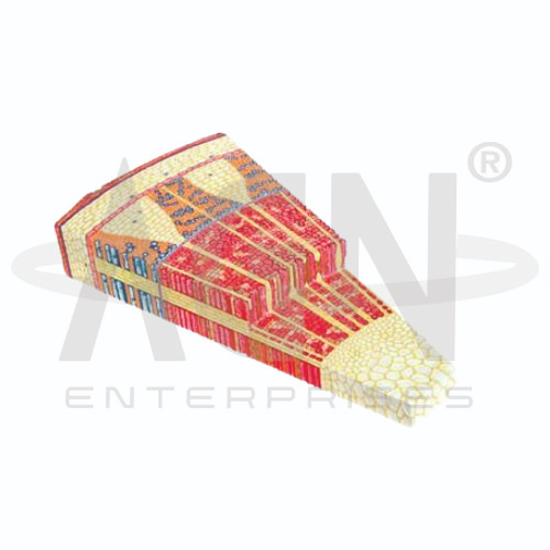
The Dicot Arboreous Stem Section (Tilia) Model from AGN Enterprises is an enlarged botanical teaching aid designed to demonstrate the internal anatomy and secondary growth of a woody dicot stem, using Tilia as a representative example. The model clearly illustrates the organization of bark, cortex, phloem, vascular cambium, secondary xylem, medullary rays, and pith.
It is suitable for schools, colleges, universities, botany laboratories, biology departments, agricultural institutes, forestry programs, horticulture departments, and plant-science training centers.
The Dicot Arboreous Stem Section (Tilia) Model provides an enlarged three-dimensional representation of the tissues found in a woody dicot stem.
Depending on the specific model configuration, represented structures may include:
Bark
Cork
Cork Cambium
Cortex
Secondary Phloem
Vascular Cambium
Secondary Xylem
Annual Growth Rings
Medullary Rays
Pith
The exact structures, colors, magnification, and sectional details may vary according to the supplied model.
Tilia is commonly used in botanical education as an example for studying secondary growth in dicotyledonous woody stems.
As the stem increases in diameter, vascular cambium and cork cambium contribute to the production of secondary tissues.
The enlarged model makes these developmental changes easier for students to visualize than microscopic sections alone.
The term bark generally refers to tissues located outside the vascular cambium in a woody stem.
These tissues provide protection and include secondary phloem and outer protective tissues.
The model helps learners understand the location of bark relative to the cambium and woody xylem.
During secondary growth, the original epidermal tissues are replaced by a protective periderm.
The cork cambium produces cork cells toward the outside and contributes to formation of the protective outer covering of the woody stem.
Where represented, students can examine the position of these tissues within the stem.
The cortex lies within the outer regions of the stem and consists largely of ground tissues.
As secondary growth progresses, the organization of the outer stem changes considerably.
The model provides a useful reference for comparing the cortical region with the vascular tissues located further inward.
The secondary phloem is produced toward the outside of the vascular cambium.
Its principal function is the transport of organic substances, including sugars produced during photosynthesis.
The model helps students identify secondary phloem and compare its position with secondary xylem.
The vascular cambium is a lateral meristem responsible for much of the increase in stem diameter during secondary growth.
It produces:
Secondary Xylem – Toward the Inside
Secondary Phloem – Toward the Outside
The model clearly demonstrates the central role of vascular cambium in woody stem development.
The secondary xylem accumulates toward the interior of the stem and forms much of the wood.
It performs important functions in water transport, mineral conduction, mechanical support, and storage.
Repeated cambial activity results in increasing quantities of secondary xylem as the stem grows.
Seasonal variations in cambial activity can produce recognizable growth rings within the secondary xylem.
These rings may reflect differences between wood produced during different parts of a growing season.
Where represented, the model provides a useful visual aid for introducing students to annual rings and woody growth.
Medullary or vascular rays extend radially through the stem.
They contribute to the lateral transport and storage of substances between inner and outer stem regions.
The model helps students visualize these radial structures in relation to the surrounding xylem and phloem.
The pith occupies the central region of the stem and consists mainly of parenchymatous tissue.
It is associated primarily with storage and represents an important landmark when studying the internal organization of a woody dicot stem.
The model helps learners compare the central pith with surrounding secondary tissues.
The model can be used to demonstrate the general sequence of secondary growth:
Vascular Cambium Activity → Secondary Xylem Formation Inward → Secondary Phloem Formation Outward → Increase in Stem Diameter
Additional activity of the cork cambium contributes to development of protective outer tissues.
This makes the model particularly valuable for studying woody plant development.
The Dicot Arboreous Stem Section (Tilia) Model provides an enlarged three-dimensional approach to studying secondary growth and woody stem anatomy.
Teachers can use it for lessons involving vascular cambium, cork cambium, secondary xylem, secondary phloem, bark, annual rings, medullary rays, pith, and secondary growth.
The model supports tissue identification, comparative plant anatomy, practical demonstrations, and examination preparation.
AGN Enterprises supplies botanical models, plant-anatomy models, biological teaching aids, scientific instruments, and educational laboratory equipment for schools, colleges, universities, agricultural institutes, forestry programs, and professional laboratories.
Our botanical teaching models provide clear three-dimensional representations to support practical botany and biology education.









