
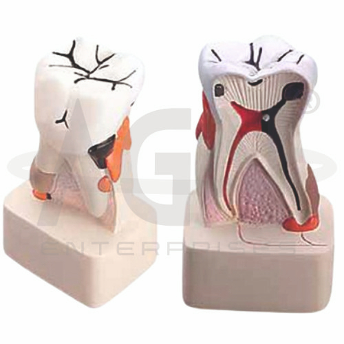
The Dental Pathology Model from AGN Enterprises is a detailed anatomical and pathological teaching aid designed to demonstrate common diseases and abnormal conditions affecting the teeth, gums, and surrounding oral structures. The enlarged three-dimensional presentation helps students compare healthy dental anatomy with selected pathological changes.
The model is suitable for dental colleges, medical colleges, nursing institutes, dental hygiene programs, pathology departments, dental clinics, oral-health centers, and professional training institutions.
The Dental Pathology Model provides an enlarged representation of normal dental structures and selected oral pathologies.
Depending on the specific model configuration, represented structures and conditions may include:
Enamel
Dentin
Dental Pulp
Gingiva
Dental Caries
Plaque and Calculus
Gingivitis
Periodontal Disease
Pulpal Involvement
Root and Supporting Structures
The exact diseases, stages, colors, sections, and anatomical details may vary according to the supplied model.
Dental caries, commonly known as tooth decay, involves progressive demineralization and destruction of tooth tissues.
The process may initially affect the enamel before extending into the dentin and, in advanced cases, approaching or involving the dental pulp.
Where represented, the model helps students visualize different depths and stages of carious lesions.
Enamel forms the highly mineralized outer covering of the anatomical crown, while dentin forms much of the underlying tooth structure.
Dental pathology can affect these tissues differently depending on the disease process and its progression.
The enlarged model makes their structural relationship easier to understand.
The dental pulp is the soft connective tissue within the central portion of the tooth and contains blood vessels and nerves.
Deep caries or other injuries may result in inflammation or infection involving pulpal tissues.
The model can help learners understand the relationship between external tooth damage and deeper pulpal involvement.
Gingivitis is inflammation of the gingiva and is commonly associated with dental plaque accumulation.
Typical clinical features can include redness, swelling, and bleeding of the gums.
Where represented, the model provides a useful visual comparison between healthy gingiva and inflammatory changes.
Periodontal disease affects the tissues that surround and support the teeth.
As periodontal disease progresses, it can involve deeper supporting structures and contribute to periodontal pocket formation, attachment loss, and alveolar bone loss.
The model can support explanations of the progression from superficial gingival inflammation to more advanced periodontal disease.
Dental plaque is a microbial biofilm that develops on tooth surfaces.
When plaque mineralizes, it can form dental calculus, also known as tartar.
Where these conditions are represented, the model helps students understand their locations and their relevance to periodontal and oral-health education.
The Dental Pathology Model can also support general teaching of normal tooth anatomy.
Important structures may include the crown, root, enamel, dentin, pulp, gingiva, periodontal tissues, and surrounding alveolar region.
Understanding normal anatomy provides an essential foundation for recognizing pathological changes.
Depending on the represented conditions, educators can use the model to explain simplified disease progressions such as:
Healthy Tooth → Plaque Accumulation → Caries → Dentin Involvement → Possible Pulpal Involvement
and
Healthy Gingiva → Gingivitis → Periodontal Disease → Supporting Tissue Damage
Actual disease progression varies according to multiple biological, behavioral, and clinical factors.
The model can support discussions about preventive oral-health measures, including:
Its enlarged format makes it particularly useful for patient and community oral-health demonstrations.
The Dental Pathology Model provides students with a three-dimensional approach to studying common dental diseases.
Teachers can use it for lessons involving dental caries, gingivitis, periodontal disease, enamel, dentin, dental pulp, plaque, calculus, oral pathology, and preventive dentistry.
The model supports pathological identification, comparison with healthy anatomy, clinical demonstrations, and examination preparation.
AGN Enterprises supplies dental models, pathological models, anatomical teaching aids, biological models, scientific instruments, and educational laboratory equipment for dental colleges, medical institutions, nursing programs, schools, hospitals, and professional training centers.
Our educational models provide clear three-dimensional representations for practical dental and medical education.









