
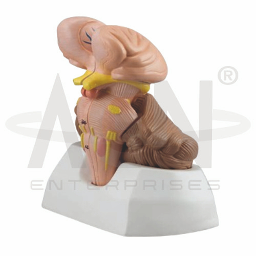
The Cerebellum and Fourth Ventricle (Brainstem) Model from AGN Enterprises is a detailed neuroanatomical teaching aid designed to demonstrate the cerebellum, fourth ventricle, brainstem, and their important anatomical relationships.
The model provides students with a three-dimensional view of structures associated with motor coordination, balance, cerebrospinal fluid circulation, and vital brainstem functions.
It is suitable for medical colleges, nursing institutes, physiotherapy colleges, neuroscience departments, paramedical institutions, anatomy laboratories, and health-science training centers.
The Cerebellum and Fourth Ventricle (Brainstem) Model demonstrates important structures of the posterior brain and brainstem region.
Depending on the specific model configuration, represented structures may include:
Cerebellum
Cerebellar Hemispheres
Vermis
Cerebellar Folia
Arbor Vitae
Fourth Ventricle
Midbrain
Pons
Medulla Oblongata
Cerebellar Peduncles
Associated Neural Structures
The exact structures, sectional details, colors, and removable components may vary according to the supplied model.
The cerebellum is located posterior to the brainstem and plays an important role in coordinating movement.
It contributes to balance, posture, motor learning, muscle coordination, and the timing and precision of voluntary movements.
The model helps students understand the position of the cerebellum relative to the brainstem and fourth ventricle.
The cerebellum consists of right and left hemispheres connected by a central region called the vermis.
The cerebellar hemispheres contribute to the coordination and refinement of voluntary movements.
The model provides a clear three-dimensional reference for studying their external organization and relationship with surrounding structures.
The vermis is the midline portion of the cerebellum connecting its two hemispheres.
It is particularly associated with the regulation of posture and coordination of movements involving the axial regions of the body.
Where represented, the model helps learners distinguish the vermis from the lateral cerebellar hemispheres.
The external surface of the cerebellum contains numerous narrow folds known as folia.
These folds increase the surface area of the cerebellar cortex.
Their characteristic arrangement can be clearly appreciated on a three-dimensional anatomical model.
The internal white matter of the cerebellum forms a branching pattern known as the arbor vitae, meaning “tree of life.”
Where the internal cerebellum is exposed, the model can help students distinguish the branching white matter from the surrounding cerebellar cortex.
The fourth ventricle is one of the interconnected cavities of the ventricular system containing cerebrospinal fluid.
It is located between the pons and upper medulla anteriorly and the cerebellum posteriorly.
The model provides an excellent three-dimensional reference for understanding this important anatomical relationship.
The fourth ventricle forms part of the ventricular pathway through which cerebrospinal fluid (CSF) circulates.
A simplified ventricular sequence is:
Lateral Ventricles → Third Ventricle → Cerebral Aqueduct → Fourth Ventricle
From the fourth ventricle, CSF can pass into spaces surrounding the brain and spinal cord.
The model helps students connect ventricular anatomy with cerebrospinal fluid circulation.
The brainstem connects higher regions of the brain with the spinal cord.
Its principal divisions are:
Midbrain
Pons
Medulla Oblongata
These regions contain important neural pathways and nuclei involved in sensory, motor, autonomic, and cranial nerve functions.
The pons forms the prominent middle portion of the brainstem and lies directly anterior to the cerebellum.
It contains important ascending and descending pathways and has extensive connections with the cerebellum.
The model helps students understand the close relationship between the pons, cerebellum, and fourth ventricle.
The medulla oblongata forms the inferior portion of the brainstem and continues into the spinal cord.
It contains important centers associated with respiratory, cardiovascular, and other autonomic functions.
The fourth ventricle is closely related to the dorsal aspect of the upper medulla.
The cerebellum communicates with the brainstem through paired fiber bundles known as the cerebellar peduncles.
These are classified as:
Superior Cerebellar Peduncles
Middle Cerebellar Peduncles
Inferior Cerebellar Peduncles
Where represented, these structures help learners understand communication between the cerebellum and other parts of the central nervous system.
The Cerebellum and Fourth Ventricle (Brainstem) Model provides a three-dimensional approach to studying complex posterior brain anatomy.
Teachers can use it for lessons involving cerebellar anatomy, fourth ventricle, brainstem, pons, medulla, cerebrospinal fluid, cerebellar peduncles, balance, and motor coordination.
It supports anatomical identification, spatial visualization, practical demonstrations, and examination preparation.
AGN Enterprises supplies neurological models, brain models, nervous-system teaching aids, anatomical models, scientific instruments, and educational laboratory equipment for medical colleges, nursing institutes, physiotherapy programs, universities, and professional training centers.
Our anatomical teaching models provide clear three-dimensional representations for practical neuroanatomy and medical education.









