
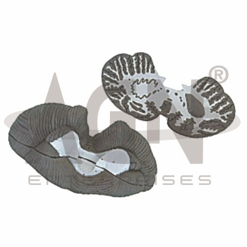
The Cerebellum 2 Parts Model from AGN Enterprises is a detailed neuroanatomical teaching aid designed to demonstrate the external and internal anatomy of the human cerebellum. Its two-part construction allows the model to be separated for closer examination of important cerebellar structures.
The model helps students understand the cerebellar hemispheres, vermis, cerebellar cortex, white matter, and associated internal structures, as represented in the supplied model.
It is suitable for medical colleges, nursing institutes, physiotherapy colleges, neuroscience departments, paramedical institutions, anatomy laboratories, and health-science training centers.
The Cerebellum 2 Parts Model provides a three-dimensional representation of the cerebellum and its characteristic anatomical organization.
Depending on the specific model configuration, represented structures may include:
Right Cerebellar Hemisphere
Left Cerebellar Hemisphere
Vermis
Cerebellar Cortex
Cerebellar Folia
White Matter
Arbor Vitae
Deep Cerebellar Structures
Cerebellar Peduncle Regions
The exact structures, colors, sectional planes, and anatomical details may vary according to the supplied model.
A key feature of this model is its two-part design.
The separable construction provides access to internal cerebellar anatomy that cannot be fully appreciated from the external surface alone.
Students can examine the external morphology and then separate the model to study represented internal structures, making it useful for practical neuroanatomy demonstrations.
The cerebellum is located in the posterior cranial fossa, behind the brainstem and inferior to the posterior regions of the cerebrum.
It plays an important role in the coordination and refinement of movement, maintenance of balance and posture, motor learning, and regulation of muscle activity.
The model helps students understand its location and characteristic three-dimensional form.
The cerebellum consists of right and left cerebellar hemispheres connected by a central region known as the vermis.
The hemispheres participate in the coordination and fine adjustment of movements.
The model provides a clear visual reference for comparing the two sides of the cerebellum.
The vermis is the narrow midline portion connecting the cerebellar hemispheres.
It has important functional relationships with the control of posture, balance, and movements involving the axial body.
Where clearly represented, the model allows students to identify the vermis relative to the lateral hemispheres.
The outer portion of the cerebellum consists of the cerebellar cortex, which is arranged into numerous narrow folds called folia.
These folds substantially increase the surface area of the cerebellar cortex.
The model’s enlarged three-dimensional representation makes the characteristic folded appearance easier to study.
Within the cerebellum lies branching white matter that forms a distinctive tree-like pattern when viewed in section.
This appearance is known as the arbor vitae, meaning “tree of life.”
Where represented, the two-part construction helps students visualize the arbor vitae and its relationship with the surrounding cerebellar cortex.
The cerebellum communicates with the brainstem through three paired groups of nerve-fiber bundles known as the cerebellar peduncles:
Superior Cerebellar Peduncles
Middle Cerebellar Peduncles
Inferior Cerebellar Peduncles
Where represented, these structures help students understand the major connections between the cerebellum and other parts of the central nervous system.
The cerebellum contributes to several important neurological functions, including:
The model helps students connect cerebellar structure with these important functions.
The cerebellum is closely associated with the midbrain, pons, and medulla oblongata through its neural connections.
Studying the cerebellum alongside a brainstem model helps learners understand how sensory and motor information is integrated for coordinated movement and balance.
The Cerebellum 2 Parts Model provides students with a three-dimensional approach to studying cerebellar anatomy.
Teachers can use it for lessons involving cerebellar hemispheres, vermis, folia, cerebellar cortex, arbor vitae, white matter, cerebellar peduncles, balance, and motor coordination.
Its separable construction supports anatomical identification, internal-structure study, practical demonstrations, and examination preparation.
AGN Enterprises supplies brain models, neurological models, nervous-system teaching aids, anatomical models, scientific instruments, and educational laboratory equipment for medical colleges, nursing institutes, physiotherapy programs, universities, and professional training centers.
Our anatomical teaching models provide clear three-dimensional representations for practical neuroanatomy and medical education.









