
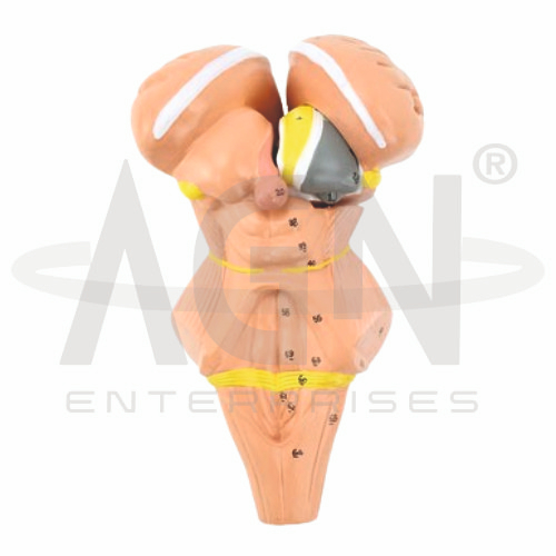
The Brainstem and Nuclei of the Hypothalamus Model from AGN Enterprises is a detailed neuroanatomical teaching aid designed to demonstrate the brainstem, hypothalamic region, and important nuclei associated with autonomic, endocrine, and homeostatic regulation.
The model provides a three-dimensional approach to complex neural anatomy, helping students understand the location and relationships of the midbrain, pons, medulla oblongata, hypothalamus, and associated neural structures.
It is suitable for medical colleges, nursing institutes, neuroscience departments, physiotherapy colleges, paramedical institutions, anatomy laboratories, psychology programs, and health-science training centers.
The Brainstem and Nuclei of the Hypothalamus Model demonstrates important structures of the central nervous system.
Depending on the specific model configuration, represented structures may include:
Midbrain
Pons
Medulla Oblongata
Hypothalamus
Hypothalamic Nuclei
Thalamic Region
Pituitary-Related Structures
Third Ventricle
Cranial Nerve Regions
Associated Neural Pathways
The exact nuclei, pathways, colors, removable components, and anatomical details may vary according to the supplied model.
The brainstem connects higher regions of the brain with the spinal cord and contains numerous pathways and nuclei involved in essential neurological functions.
Its three principal divisions are:
Midbrain
Pons
Medulla Oblongata
The model helps students visualize their arrangement and relationships with adjacent regions of the brain.
The midbrain, or mesencephalon, forms the superior portion of the brainstem.
It contains important neural structures involved in motor control, sensory processing, eye movements, visual and auditory reflexes, and communication between different regions of the central nervous system.
The model provides a useful three-dimensional reference for studying its location.
The pons lies between the midbrain and medulla oblongata.
It contains ascending and descending neural pathways and nuclei associated with several cranial nerves. It also participates in functions involving respiration, sleep, facial sensation, and motor control.
The model helps learners understand its position within the brainstem.
The medulla oblongata forms the inferior portion of the brainstem and continues into the spinal cord.
It contains neural centers involved in essential autonomic functions such as cardiovascular and respiratory regulation.
The model helps students appreciate the medulla’s important relationship with both the brain and spinal cord.
The hypothalamus is a relatively small but functionally important region of the diencephalon located inferior to the thalamus.
It plays a major role in maintaining homeostasis and contributes to the regulation of:
The hypothalamus contains multiple specialized groups of neurons known as hypothalamic nuclei.
Depending on the educational model, represented nuclei may include structures such as the:
Supraoptic Nucleus
Paraventricular Nucleus
Suprachiasmatic Nucleus
Arcuate Nucleus
Ventromedial Nucleus
Dorsomedial Nucleus
Anterior Hypothalamic Region
Posterior Hypothalamic Region
The exact nuclei displayed should be confirmed according to the supplied model.
The supraoptic and paraventricular nuclei contain neurosecretory neurons with important roles in hypothalamic-pituitary regulation.
These nuclei are associated with hormones including vasopressin (antidiuretic hormone) and oxytocin, which are transported to and released from the posterior pituitary.
Where represented, the model helps connect neural anatomy with endocrine physiology.
The suprachiasmatic nucleus is an important regulator of circadian rhythms.
It receives information related to environmental light and contributes to coordination of the body’s daily biological rhythms.
Its representation can support lessons involving sleep, biological clocks, and neuroendocrine physiology.
The hypothalamus has a close functional relationship with the pituitary gland.
Through neural and hormonal mechanisms, the hypothalamus regulates numerous endocrine functions involving growth, metabolism, reproduction, stress responses, and water balance.
Where represented, the model helps students visualize this important neuroendocrine connection.
The hypothalamus plays a central role in coordinating the autonomic nervous system.
Through connections with the brainstem and other neural regions, it contributes to regulation of cardiovascular activity, digestion, thermoregulation, and other involuntary physiological processes.
The model is therefore useful for integrating neuroanatomy with autonomic physiology.
The Brainstem and Nuclei of the Hypothalamus Model provides a three-dimensional approach to studying complex central nervous system anatomy.
Teachers can use it for lessons involving brainstem anatomy, hypothalamic nuclei, autonomic regulation, endocrine control, homeostasis, cranial nerve regions, neurophysiology, and neuroscience.
It supports anatomical identification, spatial visualization, practical demonstrations, and examination preparation.
AGN Enterprises supplies neurological models, brain models, nervous-system models, anatomical teaching aids, scientific instruments, and educational laboratory equipment for medical colleges, nursing institutes, neuroscience departments, universities, and professional training centers.
Our anatomical teaching models provide clear three-dimensional representations for practical neuroanatomy and medical education.









