
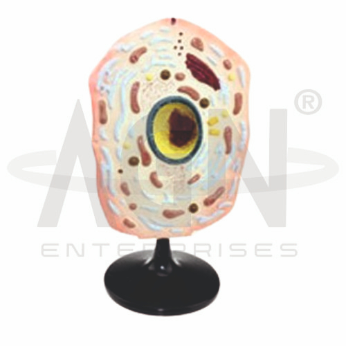
The Animal Cell Model from AGN Enterprises is an enlarged three-dimensional biological teaching aid designed to demonstrate the internal structure and major organelles of a typical animal cell. The model makes microscopic cellular structures easier to identify and understand during biology and cell-science lessons.
It provides a visual representation of important components such as the cell membrane, nucleus, nucleolus, cytoplasm, mitochondria, endoplasmic reticulum, Golgi apparatus, ribosomes, lysosomes, and other cellular structures.
The model is suitable for schools, colleges, universities, medical institutions, biology laboratories, zoology departments, biotechnology programs, and life-science training centers.
The Animal Cell Model provides an enlarged representation of the internal organization of a generalized eukaryotic animal cell.
Depending on the specific model configuration, represented structures may include:
Cell Membrane
Cytoplasm
Nucleus
Nucleolus
Nuclear Envelope
Mitochondria
Rough Endoplasmic Reticulum
Smooth Endoplasmic Reticulum
Golgi Apparatus
Ribosomes
Lysosomes
Centrosomal Structures
Vesicles
The exact organelles, colors, magnification, and structural details may vary according to the supplied model.
The cell membrane, or plasma membrane, forms the outer boundary of an animal cell.
It separates the intracellular environment from the surroundings and selectively regulates the movement of substances into and out of the cell.
The model helps students understand the membrane’s position relative to the cytoplasm and internal organelles.
The cytoplasm occupies much of the interior of the cell and surrounds the organelles.
It includes the cytosol and cellular components involved in numerous metabolic and biochemical processes.
The enlarged model provides a clear visual representation of how different organelles are distributed throughout the cell.
The nucleus is a prominent organelle in most animal cells and contains most of the cell’s genetic material.
It plays a central role in regulating gene expression and cellular activity.
The model clearly demonstrates the nucleus and its position relative to the surrounding cytoplasm.
The nucleolus is a dense region located within the nucleus.
It is involved in the production of ribosomal RNA and assembly of ribosomal subunits.
Its distinctive position within the nucleus makes it an important structure for basic cell-biology education.
The nucleus is surrounded by a nuclear envelope, consisting of a double membrane that separates nuclear contents from the cytoplasm.
Nuclear pores regulate molecular transport between these regions.
Where represented, the model helps learners distinguish the nuclear boundary from other cellular membranes.
Mitochondria are membrane-bound organelles involved in aerobic cellular respiration and ATP production.
Their internal membranes form folds known as cristae, which increase the surface area available for important metabolic reactions.
The Animal Cell Model helps students locate mitochondria and understand their role in cellular energy metabolism.
The endoplasmic reticulum (ER) is an interconnected membrane system within the cell.
It is commonly divided into:
Rough Endoplasmic Reticulum – associated with ribosomes and protein synthesis
Smooth Endoplasmic Reticulum – involved in lipid synthesis and other metabolic functions
Where represented, students can compare these two regions using the model.
The Golgi apparatus consists of stacks of flattened membrane-bound compartments.
It modifies, sorts, and packages proteins and lipids for transport to different cellular destinations.
The model provides a useful three-dimensional reference for identifying the Golgi apparatus among other cytoplasmic organelles.
Ribosomes are cellular structures responsible for protein synthesis.
They may occur freely within the cytoplasm or be associated with the rough endoplasmic reticulum.
Where represented, the model helps students understand their relationship with protein production and the endomembrane system.
Lysosomes contain enzymes involved in intracellular digestion and recycling of cellular material.
Various vesicles transport and store substances within the cell.
Where represented, these structures help students understand intracellular transport and cellular waste-processing mechanisms.
The model is useful for comparing animal cells with plant cells.
Unlike a typical plant cell, a typical animal cell does not possess a cellulose cell wall, chloroplasts, or a large central vacuole.
This makes the Animal Cell Model useful for comparative cell-biology lessons alongside plant-cell and chloroplast models.
The Animal Cell Model provides an enlarged three-dimensional approach to studying eukaryotic cell structure.
Teachers can use it for lessons involving cell organelles, nucleus, mitochondria, endoplasmic reticulum, Golgi apparatus, ribosomes, lysosomes, cell membrane, and cytoplasm.
It supports organelle identification, comparative cell biology, classroom demonstrations, practical learning, and examination preparation.
AGN Enterprises supplies cell models, biological models, botanical models, microbiology models, anatomical teaching aids, scientific instruments, and educational laboratory equipment for schools, colleges, universities, medical institutions, and professional laboratories.
Our educational models provide clear three-dimensional representations for practical biology and life-science education.









