
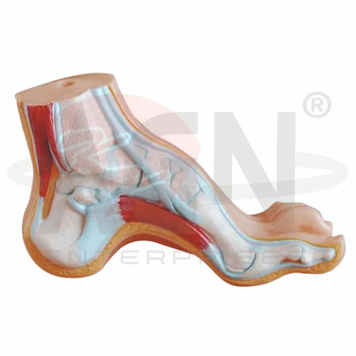
The Arched Foot Model from AGN Enterprises is a detailed anatomical teaching aid designed to demonstrate the normal arches of the human foot and the relationship between bones, ligaments, tendons, muscles, and surrounding structures.
The model provides a three-dimensional view of normal foot architecture, making it particularly useful for comparison with conditions such as flat foot (pes planus). It helps students understand how the arches contribute to weight distribution, balance, shock absorption, and efficient movement.
It is suitable for medical colleges, nursing institutes, physiotherapy colleges, orthopedic departments, podiatry programs, anatomy laboratories, paramedical institutions, and health-science training centers.
The Arched Foot Model demonstrates the anatomical organization of a normally arched human foot.
Depending on the specific model configuration, represented structures may include:
Tarsal Bones
Metatarsal Bones
Phalanges
Talus
Calcaneus
Foot Joints
Ligaments
Tendons
Muscular Structures
Plantar Region
Longitudinal Arch
The exact structures, colors, dimensions, sectional details, and anatomical components may vary according to the supplied model.
The human foot has a specialized arrangement of bones and supporting soft tissues that creates a system of anatomical arches.
These arches help the foot support body weight, absorb mechanical forces, adapt to surfaces, and provide leverage during walking and running.
The model provides a useful three-dimensional reference for studying normal foot architecture.
The medial longitudinal arch is the higher and more prominent longitudinal arch of the foot.
It is formed by several bones, including the calcaneus, talus, navicular, cuneiforms, and medial metatarsals.
Its structure and supporting tissues contribute significantly to shock absorption and efficient locomotion.
The lateral longitudinal arch is lower and flatter than the medial arch.
It is formed primarily by the calcaneus, cuboid, and lateral metatarsals.
This arch contributes to stability and helps support the foot during standing and movement.
The transverse arch extends across the foot and is associated with the arrangement of the tarsal and metatarsal regions.
Together with the longitudinal arches, it contributes to the characteristic three-dimensional shape of the foot.
The model can help students understand how these arches work together as a functional system.
The human foot normally contains 26 bones, organized into:
7 Tarsal Bones
5 Metatarsal Bones
14 Phalanges
Their arrangement forms the structural foundation of the foot and contributes to the maintenance of its normal arches.
The talus transfers forces from the lower leg into the foot and participates in the ankle joint.
The calcaneus is the largest tarsal bone and forms the heel.
Both bones are important components of the hindfoot and contribute to the mechanical organization of the foot arches.
The arches of the foot are supported by the shape of the bones as well as by ligaments, plantar connective tissues, tendons, and muscles.
Where represented, these structures help students understand that normal arch maintenance depends on both skeletal and soft-tissue components.
The Arched Foot Model is particularly useful when studied alongside a Flat Foot Model.
A normal arched foot demonstrates the characteristic elevation of the longitudinal arch, whereas flat foot or pes planus involves a reduction in the height of the medial longitudinal arch.
Comparing both models helps students recognize structural differences in foot alignment.
The arches contribute to the distribution of forces across the foot during standing and movement.
They also provide flexibility and help absorb mechanical impact during walking, running, and other weight-bearing activities.
The model helps connect anatomical structure with practical foot biomechanics.
The Arched Foot Model provides students with a three-dimensional approach to studying normal foot anatomy and biomechanics.
Teachers can use it for lessons involving foot arches, tarsal bones, metatarsals, ligaments, tendons, weight bearing, gait, flat foot comparison, and lower-limb biomechanics.
It supports anatomical identification, comparative study, practical demonstrations, and examination preparation.
AGN Enterprises supplies foot models, skeletal models, joint models, anatomical teaching aids, scientific instruments, and educational laboratory equipment for medical colleges, nursing institutes, physiotherapy programs, orthopedic departments, universities, and professional training centers.
Our anatomical models provide clear three-dimensional representations for practical musculoskeletal and human anatomy education.









