
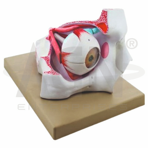
The Human Eye in Bony Orbit Model from AGN Enterprises is a detailed anatomical teaching model designed to demonstrate the structure of the human eye and its anatomical relationship with the surrounding bony orbit. It provides a clear three-dimensional view of important ocular and orbital structures involved in vision, eye movement, and protection of the eyeball.
The model is suitable for medical colleges, nursing institutes, optometry colleges, ophthalmology departments, paramedical institutions, anatomy laboratories, biology departments, and health-science training centers.
The Human Eye in Bony Orbit Model demonstrates the position of the eyeball within the orbital cavity and its relationship with surrounding structures.
Depending on the specific model configuration, represented structures may include:
Bony Orbit
Eyeball
Cornea
Sclera
Iris
Pupil
Lens
Retina
Choroid
Optic Nerve
Extraocular Muscles
Associated Nerves and Blood Vessels
The exact structures, colors, magnification, removable components, and anatomical details may vary according to the supplied model.
The bony orbit is the cavity of the skull that contains and protects the eyeball.
In addition to the globe itself, the orbit accommodates structures such as extraocular muscles, nerves, blood vessels, connective tissues, and associated ocular structures.
The model helps students understand the position of the eye within this protective skeletal cavity.
The eyeball is the principal sensory organ responsible for vision.
Its specialized tissues receive incoming light, focus it onto the retina, and initiate neural signals that are transmitted toward the brain.
The model provides a three-dimensional reference for studying the eyeball within its normal orbital environment.
The cornea forms the transparent anterior portion of the fibrous outer coat of the eye and plays an important role in refracting incoming light.
The sclera forms the tough, opaque portion of the outer coat and contributes to protection and maintenance of the shape of the eyeball.
The model helps learners compare these anatomically continuous but functionally different regions.
The iris is the colored structure located anterior to the lens.
At its center is the pupil, an opening through which light enters the deeper structures of the eye.
Changes in pupil diameter help regulate the amount of light entering the eye.
The lens is a transparent structure situated behind the iris.
Together with the cornea, it helps focus incoming light onto the retina. Changes in lens shape contribute to accommodation when focusing on objects at different distances.
Where removable or sectioned, the model allows its anatomical position to be examined more clearly.
The retina is the light-sensitive neural layer located within the posterior portion of the eyeball.
It contains specialized photoreceptors that respond to light and initiate signals involved in visual perception.
The model helps students understand the position of the retina relative to the choroid, sclera, lens, and optic nerve.
The optic nerve (cranial nerve II) carries visual information from the retina toward the brain.
It exits the posterior portion of the eyeball and passes through the orbit toward intracranial visual pathways.
Where represented, the model clearly demonstrates its relationship with the globe and surrounding orbital structures.
The position and movement of the eyeball are controlled by a coordinated group of extraocular muscles.
Where represented, these muscles help students understand how the eye moves within the bony orbit.
Their anatomical arrangement is particularly important for ophthalmology, optometry, and neuroanatomy education.
The model can support a simplified explanation of the pathway of light and visual information:
Light → Cornea → Pupil → Lens → Retina → Optic Nerve → Brain
This helps students connect the physical structures of the eye with the basic physiological process of vision.
The Human Eye in Bony Orbit Model provides students with a three-dimensional approach to studying ocular and orbital anatomy.
Teachers can use it for lessons involving bony orbit anatomy, eyeball structure, cornea, iris, lens, retina, optic nerve, extraocular muscles, ophthalmology, optometry, and vision.
It supports anatomical identification, spatial visualization, practical demonstrations, and examination preparation.
AGN Enterprises supplies anatomical models, eye models, sensory-organ models, neurological teaching aids, scientific instruments, and educational laboratory equipment for medical colleges, nursing institutes, optometry programs, universities, and professional training centers.
Our anatomical models provide clear three-dimensional representations for practical and classroom-based human anatomy education.









