
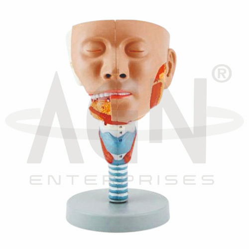
The Head with Pharynx Muscles Model from AGN Enterprises is a detailed anatomical teaching aid designed to demonstrate the muscular anatomy of the human head, pharynx, oral region, and upper neck. The model helps students understand the arrangement and relationships of muscles involved in swallowing, speech, mastication, and movement of the pharyngeal region.
It is suitable for medical colleges, dental colleges, nursing institutes, physiotherapy colleges, speech and language programs, paramedical institutions, anatomy laboratories, and health-science training centers.
The Head with Pharynx Muscles Model provides a three-dimensional representation of important structures associated with the head, throat, and pharyngeal region.
Depending on the specific model configuration, represented structures may include:
Pharyngeal Muscles
Superior Pharyngeal Constrictor
Middle Pharyngeal Constrictor
Inferior Pharyngeal Constrictor
Muscles of Mastication
Tongue-Associated Muscles
Soft Palate Region
Hyoid-Associated Muscles
Laryngeal Region
Neck Muscles
The exact structures, removable components, colors, and anatomical details may vary according to the supplied model.
The pharynx is a muscular passage extending from the region behind the nasal and oral cavities toward the esophagus and laryngeal region.
It is commonly divided into:
Nasopharynx
Oropharynx
Laryngopharynx
The model helps students understand the location and organization of these regions and their relationships with surrounding head and neck structures.
Three paired constrictor muscles form an important portion of the muscular wall of the pharynx:
Superior Pharyngeal Constrictor
Middle Pharyngeal Constrictor
Inferior Pharyngeal Constrictor
These muscles participate in coordinated movement of material through the pharynx during swallowing.
Where represented, the model makes their overlapping anatomical arrangement easier to understand.
Swallowing requires coordinated activity of muscles associated with the tongue, soft palate, pharynx, hyoid region, and larynx.
The model provides a useful three-dimensional reference for explaining how these structures interact during the swallowing process.
This makes it particularly useful for medical, dental, nursing, and speech-related education.
The oral cavity communicates posteriorly with the oropharyngeal region.
The Head with Pharynx Muscles Model helps students visualize the anatomical relationship between the mouth, tongue, palate, pharynx, and upper digestive passage.
This relationship is important for understanding mastication, swallowing, speech, and airway anatomy.
The hyoid bone provides attachment for several muscles associated with the tongue, pharynx, and laryngeal region.
The suprahyoid and infrahyoid muscles contribute to movements associated with swallowing and stabilization of the hyoid and larynx.
Where represented, these structures provide additional context for functional head-and-neck anatomy.
The pharynx forms part of both the respiratory and digestive pathways.
Air travels through the pharyngeal region toward the larynx and lower respiratory tract, while swallowed material is directed toward the esophagus.
The model helps learners understand how these pathways are anatomically related.
The Head with Pharynx Muscles Model provides students with a three-dimensional approach to studying complex head, throat, and neck anatomy.
Teachers can use it for lessons involving pharyngeal muscles, swallowing, oral anatomy, head-and-neck musculature, hyoid anatomy, upper digestive pathways, respiratory anatomy, and functional anatomy.
It supports structural identification, spatial visualization, practical demonstrations, and examination preparation.
AGN Enterprises supplies anatomical models, muscular models, head-and-neck models, skeletal teaching aids, biological models, and educational laboratory equipment for medical colleges, dental colleges, nursing institutes, physiotherapy programs, and professional training centers.
Our anatomical teaching models provide clear three-dimensional representations for practical and classroom-based human anatomy education.









