
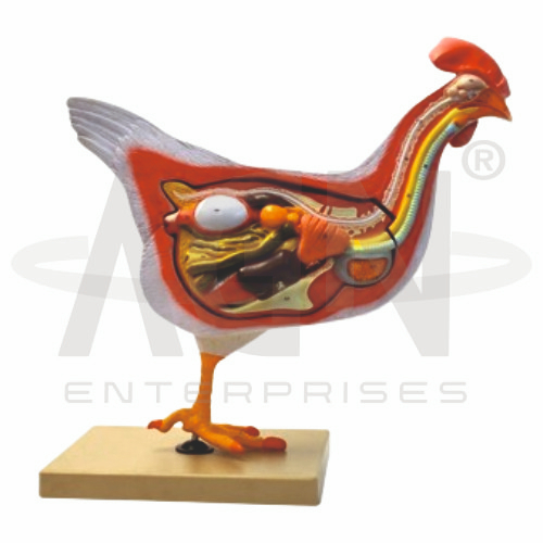
The Hen Dissection Model from AGN Enterprises is a detailed zoological teaching aid designed to demonstrate the external and internal anatomy of a hen. The model provides a three-dimensional view of major organs and body systems, helping students understand avian anatomy without depending entirely on preserved specimens or actual dissection.
It is suitable for schools, colleges, universities, zoology laboratories, biology departments, veterinary education, agricultural institutes, and educational training centers.
The Hen Dissection Model provides a clear representation of important anatomical structures and their positions within the body.
Depending on the specific model configuration, structures represented may include:
Heart
Lungs
Trachea
Esophagus
Crop
Proventriculus
Gizzard
Liver
Intestines
Kidneys
Reproductive Organs
Associated Internal Structures
The exact organs, detachable components, colors, and anatomical details may vary according to the supplied model.
The digestive system of a hen is adapted for processing food without teeth. The model can help students trace the general digestive pathway:
Mouth → Esophagus → Crop → Proventriculus → Gizzard → Small Intestine → Large Intestine → Cloaca
The crop temporarily stores food, while the glandular proventriculus and muscular gizzard contribute to digestion and mechanical breakdown.
The avian respiratory system differs significantly from that of mammals.
The model can be used to identify structures such as the trachea and lungs, along with other respiratory structures where represented. Birds also possess air sacs that participate in their highly efficient respiratory system.
This makes the model useful for comparative vertebrate anatomy.
Like mammals, birds possess a four-chambered heart consisting of two atria and two ventricles.
The heart maintains separation between oxygenated and deoxygenated blood and supports the high metabolic demands associated with avian activity.
Where represented, students can study the position of the heart relative to surrounding organs.
The Hen Dissection Model may demonstrate important structures of the female avian reproductive system.
In adult hens, the left ovary and left oviduct are normally the primary functional reproductive structures. The oviduct contains specialized regions involved in egg formation and transport.
The model helps connect reproductive anatomy with poultry and avian biology.
The kidneys of birds are positioned dorsally within the body cavity and play an important role in waste removal and regulation of internal fluid balance.
Birds excrete nitrogenous waste primarily in the form of uric acid.
Where represented, the model helps students identify the kidneys and understand their anatomical location.
The Hen Dissection Model provides a reusable alternative for introducing structures commonly examined during zoological dissection.
Students can observe the spatial relationships between the digestive, respiratory, circulatory, reproductive, and excretory systems.
This makes it particularly useful before or alongside practical dissection classes.
The model supports lessons involving hen anatomy, avian digestive system, respiratory system, circulatory system, reproductive system, excretory system, vertebrate zoology, and comparative anatomy.
Its three-dimensional construction helps students identify organs, understand their positions, compare body systems, and prepare for practical examinations.
AGN Enterprises supplies zoological models, biological teaching aids, anatomical models, botanical models, scientific instruments, and educational laboratory equipment for schools, colleges, universities, veterinary institutions, and professional laboratories.
Our teaching models provide clear three-dimensional representations to support practical biology and zoology education.









