
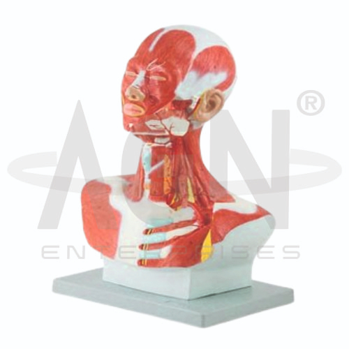
The Head and Neck Musculature Model from AGN Enterprises is a detailed anatomical teaching aid designed to demonstrate the muscles of the human head, face, and neck along with their anatomical relationships. The three-dimensional model helps students identify superficial and deeper muscular structures involved in facial expression, mastication, head movement, swallowing, and neck stabilization.
It is suitable for medical colleges, dental colleges, nursing institutes, physiotherapy colleges, paramedical institutions, anatomy laboratories, occupational therapy programs, and health-science training centers.
The Head and Neck Musculature Model provides a three-dimensional representation of important muscular structures in the craniofacial and cervical regions.
Depending on the specific model configuration, represented structures may include:
Facial Muscles
Muscles of Mastication
Temporal Muscle
Masseter Muscle
Orbicularis Oculi
Orbicularis Oris
Sternocleidomastoid
Suprahyoid Muscles
Infrahyoid Muscles
Posterior Neck Muscles
The exact structures, removable components, colors, and anatomical details may vary according to the supplied model.
The muscles of facial expression are primarily responsible for movements of the skin and soft tissues of the face.
Important muscles may include the orbicularis oculi, orbicularis oris, buccinator, frontalis, and other facial muscles.
Where represented, the model helps students understand their positions and relationships with surrounding facial structures.
The principal muscles of mastication contribute to movements of the mandible during chewing.
They include:
Masseter
Temporalis
Medial Pterygoid
Lateral Pterygoid
The model can help students understand their anatomical positions and their relationship with the skull and mandible.
The sternocleidomastoid is one of the most prominent superficial muscles of the neck.
It contributes to movements involving rotation and flexion of the head and neck. Its location also makes it an important anatomical landmark in the cervical region.
The model provides a clear three-dimensional reference for identifying this muscle.
The suprahyoid and infrahyoid muscle groups are associated with the hyoid bone, swallowing, and movements of the laryngeal region.
Where represented, these muscles help students understand the functional relationship between the floor of the mouth, hyoid bone, larynx, and anterior neck.
Muscles located in the posterior cervical region contribute to extension, rotation, stabilization, and postural control of the head and neck.
The model can help learners understand the layered arrangement of these muscles and their relationships with the skull and cervical vertebral column.
The Head and Neck Musculature Model can support demonstrations of muscular functions associated with:
This makes the model particularly useful for anatomy, dentistry, physiotherapy, and rehabilitation education.
The model provides students with a three-dimensional approach to studying the complex muscular anatomy of the head and neck.
Teachers can use it for lessons involving facial muscles, muscles of mastication, cervical muscles, hyoid muscles, functional anatomy, musculoskeletal anatomy, dentistry, and physiotherapy.
It supports muscle identification, spatial visualization, practical demonstrations, and examination preparation.
AGN Enterprises supplies anatomical models, muscular models, skeletal teaching aids, head-and-neck models, scientific instruments, and educational laboratory equipment for medical colleges, dental colleges, nursing institutes, physiotherapy programs, and professional training centers.
Our anatomical teaching models provide clear three-dimensional representations for practical and classroom-based human anatomy education.









