
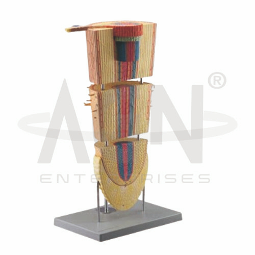
The Dicot Root Section (Leguminosae) Model from AGN Enterprises is an enlarged botanical teaching aid designed to demonstrate the internal anatomy and tissue arrangement of a typical dicotyledonous root belonging to the family Leguminosae (Fabaceae). The model provides a clear three-dimensional representation of important root tissues and vascular structures.
It is suitable for schools, colleges, universities, botany laboratories, biology departments, agricultural institutes, horticulture programs, and plant-science training centers.
The Dicot Root Section (Leguminosae) Model demonstrates the characteristic organization of tissues visible in a transverse section of a dicot root.
Depending on the specific model configuration, represented structures may include:
Epiblema / Epidermis
Root Hairs
Cortex
Endodermis
Pericycle
Phloem
Xylem
Conjunctive Tissue
Vascular Region
Central Region
The exact structures, magnification, colors, and sectional details may vary according to the supplied model.
The outermost layer of a young root is commonly referred to as the epiblema, rhizodermis, or root epidermis.
Root hairs arise from specialized epidermal cells and substantially increase the surface area available for the absorption of water and dissolved mineral nutrients from the soil.
Where represented, the enlarged model makes these structures easier to identify.
The cortex occupies a substantial region between the outer epidermal layer and the central vascular cylinder.
It consists largely of parenchymatous cells and participates in storage and movement of water and dissolved substances toward the inner tissues.
The model helps students understand the relative thickness and position of the cortical region.
The endodermis forms the innermost layer of the cortex and surrounds the vascular cylinder.
It plays an important role in regulating the movement of water and dissolved substances into the vascular tissues.
The enlarged sectional model helps learners locate the endodermis relative to the cortex and pericycle.
The pericycle is situated immediately inside the endodermis.
It is particularly important because lateral roots originate from the pericycle in typical vascular plants.
In dicot roots undergoing secondary growth, pericycle-associated tissues also contribute to subsequent developmental changes.
The xylem transports water and dissolved mineral nutrients from the root toward the aerial portions of the plant.
In a typical young dicot root, the primary xylem commonly forms a central, radial arrangement with projecting arms.
The model helps students recognize this characteristic vascular organization.
The phloem transports organic substances, particularly products of photosynthesis, throughout the plant.
In the primary dicot root, phloem strands are positioned between the arms of the xylem.
The model provides a clear visual reference for comparing xylem and phloem distribution.
A characteristic feature of root anatomy is the radial arrangement of vascular tissues, in which xylem and phloem occur on different radii.
This differs from the conjoint vascular bundles commonly encountered in stems.
The model is therefore particularly useful for comparative lessons involving root and stem anatomy.
Important features commonly associated with a young dicot root include:
These features help distinguish typical dicot root anatomy from monocot root organization.
Leguminosae, now commonly referred to as Fabaceae, includes many economically important plants such as peas, beans, lentils, and other legumes.
Many members of this family form specialized root nodules through associations with nitrogen-fixing bacteria. However, nodules and their internal structures should only be considered part of this model where specifically represented.
The Dicot Root Section (Leguminosae) Model provides an enlarged three-dimensional approach to studying plant anatomy.
Teachers can use it for lessons involving dicot root structure, epidermis, cortex, endodermis, pericycle, xylem, phloem, vascular organization, root histology, and comparative plant anatomy.
The model supports tissue identification, practical demonstrations, botanical laboratory study, and examination preparation.
AGN Enterprises supplies botanical models, plant-anatomy models, biological teaching aids, scientific instruments, and educational laboratory equipment for schools, colleges, universities, agricultural institutes, and professional laboratories.
Our botanical teaching models provide clear three-dimensional representations to support practical botany and biology education.









