
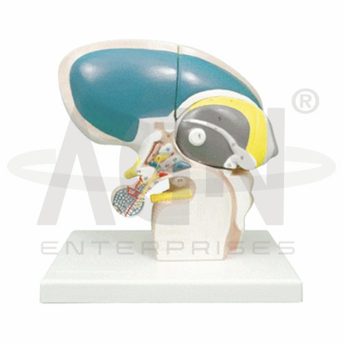
The Diencephalon Model from AGN Enterprises is a detailed anatomical teaching aid designed to demonstrate the major structures of the diencephalon and their relationships within the human brain. The model helps students study important regions such as the thalamus, hypothalamus, epithalamus, and surrounding neural structures in a clear three-dimensional format.
It is suitable for medical colleges, nursing institutes, physiotherapy colleges, neuroscience departments, paramedical institutions, psychology programs, anatomy laboratories, and health-science training centers.
The Diencephalon Model provides a three-dimensional representation of structures located deep within the brain.
Depending on the specific model configuration, represented structures may include:
Thalamus
Hypothalamus
Epithalamus
Subthalamic Region
Pineal Gland
Third Ventricle
Optic Region
Pituitary-Related Structures
Associated Neural Pathways
The exact structures, removable components, colors, and anatomical details may vary according to the supplied model.
The diencephalon is a central region of the forebrain situated between the cerebral hemispheres and the midbrain.
It contains several important structures involved in sensory processing, autonomic regulation, endocrine control, sleep, consciousness, temperature regulation, and homeostasis.
The model provides a useful three-dimensional reference for understanding this complex region.
The thalamus forms a major portion of the diencephalon.
It serves as an important relay and processing center for much of the sensory information traveling toward the cerebral cortex and also participates in motor and cognitive functions.
Where represented, the model helps students understand its central position within the brain.
The hypothalamus lies inferior to the thalamus and plays a major role in maintaining physiological homeostasis.
Its functions include regulation of:
Body Temperature
Hunger and Thirst
Autonomic Activity
Sleep-Wake Functions
Endocrine Activity
Fluid Balance
The model helps connect hypothalamic anatomy with these important physiological functions.
The epithalamus forms part of the dorsal diencephalic region and includes structures associated with the pineal gland and habenular region.
The pineal gland secretes melatonin and contributes to regulation of circadian rhythms.
Where represented, these structures help students understand the relationship between neuroanatomy and biological rhythms.
The third ventricle is a narrow cerebrospinal-fluid-filled cavity located in the midline of the diencephalon.
Its walls are closely related to the thalamic and hypothalamic regions.
The model can help learners visualize the position of the third ventricle within the ventricular system of the brain.
The hypothalamus has an important functional relationship with the pituitary gland and plays a central role in neuroendocrine regulation.
Through neural and hormonal mechanisms, the hypothalamic-pituitary system influences numerous endocrine functions throughout the body.
Where represented, the model provides useful context for studying this relationship.
The Diencephalon Model can support teaching about several important neurological and physiological functions, including:
The model provides students with a three-dimensional approach to studying deep brain anatomy.
Teachers can use it for lessons involving diencephalon anatomy, thalamus, hypothalamus, epithalamus, third ventricle, neuroendocrine regulation, autonomic functions, and neuroscience.
It supports anatomical identification, spatial visualization, practical demonstrations, and examination preparation.
AGN Enterprises supplies neurological models, brain models, anatomical teaching aids, nervous-system models, scientific instruments, and educational laboratory equipment for medical colleges, nursing institutes, physiotherapy programs, universities, and professional training centers.
Our anatomical teaching models provide clear three-dimensional representations for practical neuroanatomy and medical education.









