
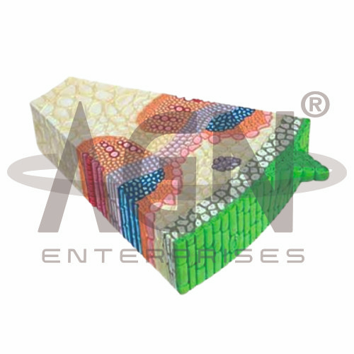
The Monocot Leaf Section (Zea Mays) from AGN Enterprises is an enlarged botanical teaching model designed to demonstrate the internal anatomy of a maize leaf. It provides a clear visual representation of important tissues such as the epidermis, mesophyll, vascular bundles, bundle sheath, xylem, phloem, stomata, and bulliform cells.
The model makes microscopic leaf structures easier to identify and understand during classroom and laboratory demonstrations. It is suitable for schools, colleges, botany laboratories, biology departments, teacher training institutes, and life-science education.
Maize (Zea mays) is commonly studied as an example of monocot leaf anatomy. A transverse section demonstrates the characteristic organization of epidermal, ground, and vascular tissues.
Students can use the model to study structures such as:
Upper Epidermis
Lower Epidermis
Mesophyll
Bulliform Cells
Vascular Bundles
Bundle Sheath
Xylem
Phloem
Stomata
The exact structures represented may vary according to the supplied model configuration.
The leaf is covered externally by epidermal tissue. The upper and lower epidermis form protective outer layers and help separate internal tissues from the external environment.
Stomata present in the epidermis participate in gas exchange and regulation of water loss.
The enlarged teaching aid makes it easier for students to understand the relationship between epidermal structures and the internal tissues of the leaf.
An important feature commonly studied in grass leaves is the presence of bulliform cells in the upper epidermal region.
These specialized epidermal cells are associated with changes in leaf folding or rolling during variations in water availability.
Their representation makes the model particularly useful when teaching characteristic features of monocot leaves.
The internal photosynthetic tissue is known as the mesophyll.
In a typical maize leaf, the mesophyll does not show the same distinct palisade and spongy differentiation commonly emphasized in many dorsiventral dicot leaves.
This feature can be used to compare monocot and dicot leaf anatomy.
The Monocot Leaf Section (Zea Mays) helps students study the vascular tissues responsible for transportation within the plant.
Xylem primarily transports water and dissolved minerals.
Phloem transports sugars and other organic substances.
Vascular bundles are surrounded by prominent bundle sheath cells, an important feature of maize leaf anatomy.
Maize is a C4 plant and exhibits characteristic Kranz anatomy.
In this arrangement, prominent bundle sheath cells surround the vascular bundles, while mesophyll cells are arranged around the bundle sheath region.
This structural specialization is associated with the C4 pathway of photosynthesis, making the maize leaf useful for teaching both plant anatomy and photosynthetic adaptations.
Teachers can use the model alongside a dicot leaf model to demonstrate important anatomical differences.
A typical maize leaf shows features such as bulliform cells, relatively similar upper and lower surfaces, undifferentiated mesophyll, parallel venation, and characteristic vascular-bundle organization.
Comparative study helps students identify structural adaptations and distinguish between common monocot and dicot leaf types.
This enlarged botanical model converts microscopic leaf anatomy into an accessible visual learning experience. Students can identify individual tissues and understand their positions and functions without depending entirely on microscope slides or textbook diagrams.
It is useful for lessons involving plant anatomy, leaf structure, monocot characteristics, vascular tissues, stomata, photosynthesis, C4 plants, and monocot-dicot comparison.
The model complements microscopy and practical botany work while supporting observation, identification, visualization, and conceptual understanding.
AGN Enterprises supplies botanical models, biology teaching aids, anatomical models, and educational laboratory equipment for schools, colleges, universities, and training institutions.
Our enlarged plant models help make microscopic botanical structures easier to demonstrate and understand during practical science education.









