
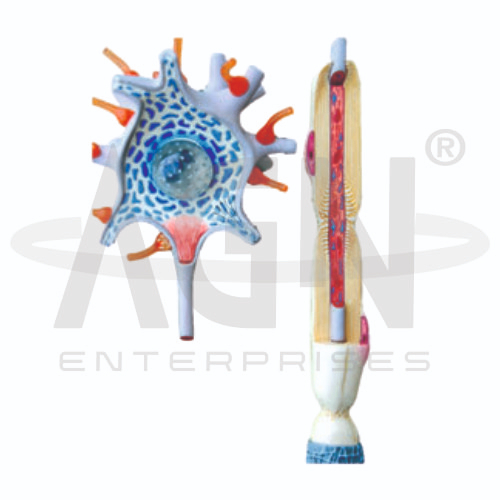
The Neuron Model from AGN Enterprises is an enlarged three-dimensional biological teaching model designed to demonstrate the structure of a neuron, the basic functional unit of the nervous system. The model helps students identify major parts such as the cell body, nucleus, dendrites, axon, myelin sheath, nodes of Ranvier, and axon terminals. It provides a clear visual aid for understanding how neurons receive and transmit signals. It is suitable for schools, colleges, medical and nursing institutes, biology laboratories, anatomy departments, and neuroscience education.
The Neuron Model provides an enlarged representation of a nerve cell, making its individual components easier to identify and study.
Depending on the specific model configuration, structures represented may include:
Dendrites
Cell Body (Soma)
Nucleus
Axon
Myelin Sheath
Nodes of Ranvier
Axon Terminals
Synaptic Endings
The three-dimensional design allows teachers to demonstrate the relationship between different parts of a neuron more clearly than with a flat diagram alone.
A neuron is a specialized cell involved in receiving, processing, and transmitting information within the nervous system.
The model can be used to explain three major regions:
Dendrites – branching structures that receive signals from other cells.
Cell Body (Soma) – contains the nucleus and supports the neuron’s cellular functions.
Axon – carries electrical signals away from the cell body toward other neurons, muscles, or glands.
This structural organization helps students understand the direction in which information generally travels through a neuron.
Where represented, the Neuron Model can demonstrate the relationship between the axon, myelin sheath, and nodes of Ranvier.
The myelin sheath forms an insulating covering around portions of many axons and helps increase the speed of electrical impulse conduction.
The gaps between myelinated segments are called the nodes of Ranvier.
These structures provide a useful foundation for explaining saltatory conduction in myelinated nerve fibers.
The Neuron Model can also support lessons on nerve-impulse transmission.
A simplified pathway can be represented as:
Dendrites → Cell Body → Axon → Axon Terminals
At the end of an axon, communication with another cell can occur across a specialized junction known as a synapse.
Teachers can use the model to introduce concepts such as electrical signaling, neurotransmission, sensory pathways, motor responses, and communication within the nervous system.
The Neuron Model transforms microscopic nervous-system anatomy into an enlarged three-dimensional learning experience. Students can identify individual neuronal structures and connect each part with its primary function.
It is useful for teaching cell biology, nervous-system anatomy, neurophysiology, nerve impulses, synapses, myelination, and neural communication.
The model complements textbooks, diagrams, microscope studies, and classroom demonstrations while improving visualization and structural understanding.
AGN Enterprises supplies anatomical models, biological models, and educational laboratory equipment for schools, colleges, medical institutions, nursing programs, and professional training centers.
Our enlarged biological models help teachers demonstrate complex structures clearly and support practical, visual, and activity-based science education.









