
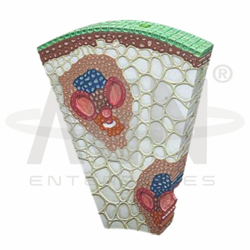
The Monocot Stem Section (Zea Mays) from AGN Enterprises is an enlarged three-dimensional botanical teaching model designed to demonstrate the internal anatomy of a monocot stem using maize (Zea mays) as a representative example. The model helps students study the arrangement of tissues such as the epidermis, ground tissue, vascular bundles, xylem, phloem, and bundle sheath in a clear and visual format. It is suitable for schools, colleges, botany laboratories, biology departments, teacher training institutes, and life-science education.
The Monocot Stem Section (Zea Mays) provides an enlarged representation of a transverse section of a maize stem, making microscopic plant tissues easier to identify during classroom demonstrations.
Depending on the specific model configuration, it may demonstrate:
Epidermis
Hypodermal Region
Ground Tissue
Vascular Bundles
Bundle Sheath
Phloem
Xylem
Protoxylem Lacuna
The enlarged model allows students to understand the characteristic internal organization of a monocot stem without relying solely on microscope slides or textbook diagrams.
The maize stem provides a commonly studied example of monocot stem anatomy.
The outer region is covered by an epidermis, while much of the internal portion consists of ground tissue.
One of the most recognizable characteristics is the presence of numerous vascular bundles scattered throughout the ground tissue, rather than arranged in a distinct ring as typically seen in many dicot stems.
This characteristic makes the model particularly useful for comparative plant-anatomy lessons.
The Monocot Stem Section (Zea Mays) can be used to study the internal structure of individual vascular bundles.
Each vascular bundle typically contains:
Phloem – involved primarily in transporting sugars and other organic substances.
Xylem – primarily conducts water and dissolved minerals.
Bundle Sheath – surrounds the vascular bundle.
In maize stems, the vascular bundles are generally described as conjoint, collateral, and closed, meaning vascular cambium is absent between the xylem and phloem within the bundle.
A characteristic feature commonly studied in maize stem anatomy is the protoxylem lacuna.
As the stem develops, some protoxylem elements can break down, leaving a cavity associated with the vascular bundle. An enlarged teaching model makes this feature easier to identify and discuss than a small textbook illustration.
The model can also support comparative lessons between monocot and dicot stems.
A typical maize monocot stem shows:
Scattered Vascular Bundles
Undifferentiated Ground Tissue
Closed Vascular Bundles
In contrast, many typical dicot stems show vascular bundles arranged in a ring and have a vascular cambium associated with secondary growth.
This comparison helps students recognize important anatomical differences between major groups of flowering plants.
The Monocot Stem Section (Zea Mays) converts microscopic plant anatomy into a large three-dimensional learning experience. Students can identify individual tissues and understand their spatial relationships more clearly.
Teachers can use the model for lessons involving plant anatomy, monocot stems, vascular tissues, xylem, phloem, vascular bundles, tissue systems, and monocot-dicot comparison.
It complements microscope slides and practical botany work while supporting observation, structural identification, visualization, and conceptual understanding.
AGN Enterprises supplies botanical models, biological teaching aids, anatomical models, and educational laboratory equipment for schools, colleges, universities, and training institutions.
Our enlarged plant-anatomy models help teachers demonstrate microscopic botanical structures clearly and support practical, visual science education.









