
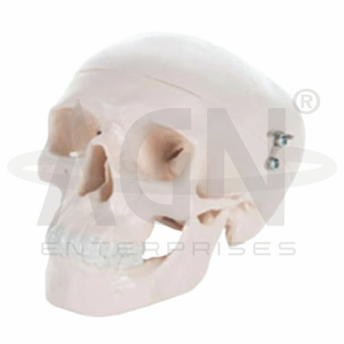
The Mini Skull Model from AGN Enterprises is a compact anatomical teaching aid designed to demonstrate the basic structure and major bones of the human skull. Its reduced-size construction provides a convenient way to study cranial anatomy while requiring less storage and display space than a full-size skull model.
The model is suitable for schools, medical colleges, nursing institutes, physiotherapy colleges, paramedical institutions, biology laboratories, anatomy departments, and health-science training centers.
The human skull forms the bony framework of the head and protects the brain while supporting structures associated with vision, hearing, breathing, and the oral cavity.
This teaching model can help students recognize major regions of the skull, including:
Cranium
Facial Skeleton
Orbits
Nasal Cavity
Upper Jaw
Lower Jaw
Cranial Base
The exact anatomical details represented depend on the supplied model configuration.
The Mini Skull Model can support identification of important cranial and facial bones, including:
Frontal Bone
Parietal Bones
Temporal Bones
Occipital Bone
Sphenoid Bone
Ethmoid Bone
Maxilla
Mandible
Zygomatic Bones
Nasal Bones
The compact three-dimensional representation helps learners understand how individual bones combine to form the skull.
The skull can broadly be studied as two groups of bones:
Cranial Bones – form the cranium and surround and protect the brain.
Facial Bones – contribute to the structure of the face, nasal region, orbits, and jaws.
Using a physical model allows students to observe the relative positions of these bones from multiple angles.
Where sufficiently detailed on the model, students can also identify the major fibrous joints between cranial bones known as sutures.
Important sutures include:
Coronal Suture
Sagittal Suture
Lambdoid Suture
Squamous Suture
These landmarks are useful in the study of osteology and head anatomy.
The reduced dimensions make this skull particularly useful where a portable and space-saving anatomical model is preferred.
Teachers can use it for individual demonstrations, student revision, anatomical identification, and comparison with diagrams or other skeletal models.
Its three-dimensional format allows learners to examine the skull from anterior, posterior, lateral, superior, and inferior perspectives, depending on the model’s construction.
This anatomical teaching aid helps students move beyond two-dimensional illustrations and develop a clearer understanding of skull structure and spatial relationships.
It is useful for teaching osteology, skeletal anatomy, cranial anatomy, facial bones, skull landmarks, and basic head anatomy.
The model can complement textbooks, anatomical charts, and classroom demonstrations while improving visual recognition and anatomical orientation.
AGN Enterprises supplies anatomical models, skeleton models, biological teaching aids, and educational laboratory equipment for schools, colleges, medical institutions, nursing programs, physiotherapy institutes, and professional training centers.
Our anatomical teaching aids are designed to provide clear visual representations for practical and classroom-based education.









