
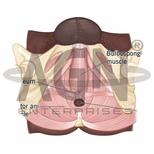
The Male Perineum Model from AGN Enterprises is a detailed anatomical teaching aid designed to demonstrate the muscles, fascia, vessels, nerves, and associated structures of the male perineal region. The model helps students understand the complex anatomical relationships between the pelvic floor, external genital region, anal region, and surrounding structures.
It is suitable for medical colleges, nursing institutes, physiotherapy colleges, paramedical institutions, anatomy laboratories, urology education, and other health-science training programs.
The perineum is the anatomical region located inferior to the pelvic floor. It is commonly described as a diamond-shaped region extending between the pubic symphysis anteriorly, coccyx posteriorly, and ischial tuberosities laterally.
For anatomical study, it is divided into two main regions:
Urogenital Triangle
Anal Triangle
This teaching model provides a three-dimensional representation that helps learners understand the arrangement of structures within these regions.
The anterior portion of the perineum forms the urogenital triangle.
Depending on the specific model configuration, structures represented may include:
Root of the Penis
Bulbospongiosus Muscle
Ischiocavernosus Muscle
Superficial Transverse Perineal Muscle
Perineal Membrane
Urethral Structures
Associated Blood Vessels and Nerves
The exact anatomical structures and level of detail may vary according to the supplied model.
The posterior portion forms the anal triangle and contains structures associated with the anal canal and pelvic floor.
The model may demonstrate structures such as the:
External Anal Sphincter
Anal Canal
Ischioanal Region
Pelvic Floor Muscles
Related Nerves and Vessels
These structures help students understand the functional relationship between the perineum, pelvic floor, and anal region.
The Male Perineum Model is particularly useful for studying the muscular organization of this anatomically complex region.
Where represented, students can identify muscles such as:
Bulbospongiosus
Ischiocavernosus
Superficial Transverse Perineal Muscle
External Anal Sphincter
Pelvic Floor Muscles
Studying these structures in three dimensions helps learners understand their positions and functional relationships.
Depending on its level of detail, the teaching aid may also demonstrate important neurovascular structures of the perineum.
The pudendal nerve and internal pudendal vessels are particularly important structures associated with this region.
Their representation supports lessons involving regional anatomy, pelvic anatomy, urology, and clinical anatomy.
The perineum has a close anatomical relationship with the pelvic floor, which supports the pelvic organs and contributes to urinary and bowel control.
Using this model, instructors can connect superficial perineal anatomy with deeper pelvic structures and explain how muscles, fascia, nerves, and vessels are organized within the region.
This anatomical teaching aid provides a clear visual approach to an area that can be difficult to understand using two-dimensional illustrations alone.
It is useful for teaching male pelvic anatomy, perineal muscles, pelvic floor anatomy, urogenital anatomy, anal anatomy, neurovascular structures, and clinical anatomy.
The model supports anatomical identification, spatial visualization, practical demonstrations, and revision for medical and allied-health students.
AGN Enterprises supplies anatomical models, biological teaching aids, pathological models, and educational laboratory equipment for medical colleges, nursing institutes, physiotherapy programs, paramedical institutions, and professional training centers.
Our anatomical models provide clear three-dimensional representations for practical demonstrations and human-anatomy education.









