
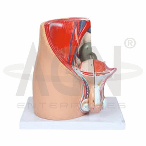
The Male Pelvis (4 Parts) Model from AGN Enterprises is a detailed anatomical teaching aid designed to demonstrate the structure of the male pelvis and the anatomical relationships of the pelvic organs. Its four-part construction allows selected components to be separated for closer examination, helping students understand male pelvic anatomy in a clear three-dimensional format.
The model is suitable for medical colleges, nursing institutes, physiotherapy colleges, paramedical institutions, anatomy laboratories, urology departments, and health-science training centers.
The male pelvis contains important structures belonging to the urinary, reproductive, and digestive systems.
Depending on the specific model configuration, structures represented may include:
Pelvic Bones
Urinary Bladder
Prostate Gland
Urethra
Rectum
Seminal Vesicles
Vas Deferens
Penis
Associated Pelvic Structures
The exact removable components and anatomical details may vary according to the supplied four-part model.
A key feature of the Male Pelvis (4 Parts) Model is its sectional, multi-part construction.
The separable components allow learners to examine deeper structures that may otherwise be difficult to observe. Students can study the location of individual organs and then understand how these structures fit together within the pelvic region.
This makes the model useful for practical demonstrations, anatomy identification, revision, and comparative study.
The urinary bladder is located within the pelvis and serves as a temporary reservoir for urine.
Immediately below the bladder lies the prostate gland, which surrounds the proximal portion of the male urethra.
The teaching model helps students visualize this important relationship and provides a useful foundation for studying male urinary anatomy, prostate anatomy, and introductory urology.
Where represented, students can examine structures associated with the male reproductive system, including the:
Seminal Vesicles
Vas Deferens
Prostate Gland
Urethra
Penile Structures
The three-dimensional presentation helps learners understand how reproductive structures are positioned relative to the bladder, rectum, and other pelvic organs.
The rectum occupies the posterior portion of the pelvic region and has important anatomical relationships with the urinary and reproductive organs.
Using the model, students can visualize the relative positions of the rectum, urinary bladder, prostate, and reproductive structures.
This spatial understanding is particularly valuable for regional anatomy and clinical education.
This anatomical teaching aid provides a practical way to study the complex arrangement of organs within the male pelvis.
Teachers can use it for lessons involving male pelvic anatomy, urinary anatomy, reproductive anatomy, prostate anatomy, urology, regional anatomy, and human anatomy.
The four-part construction supports hands-on learning while improving anatomical identification, spatial visualization, and understanding of organ relationships.
AGN Enterprises supplies anatomical models, biological teaching aids, pathological models, and educational laboratory equipment for medical colleges, nursing institutes, physiotherapy programs, paramedical institutions, schools, and professional training centers.
Our anatomical models provide clear three-dimensional representations for classroom demonstrations and practical human-anatomy education.









