
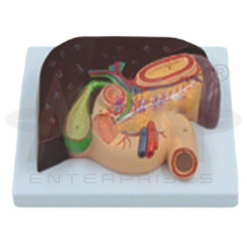
The Liver & Gallbladder (Pancreas & Duodenum) Model from AGN Enterprises is a detailed anatomical teaching aid designed to demonstrate the liver, gallbladder, pancreas, duodenum, and their associated duct systems. The model provides a three-dimensional representation of these closely related digestive organs, helping students understand their positions, structural relationships, and roles in digestion.
It is suitable for medical colleges, nursing institutes, paramedical institutions, biology laboratories, anatomy departments, gastroenterology education, and health-science training centers.
The liver is a major organ involved in metabolism, nutrient processing, storage, detoxification, and bile production. The gallbladder is closely associated with the liver and stores and concentrates bile.
Depending on the specific model configuration, structures represented may include:
Liver
Right and Left Lobes
Gallbladder
Hepatic Ducts
Cystic Duct
Common Bile Duct
Pancreas
Pancreatic Duct
Duodenum
Associated Blood Vessels
The exact anatomical details may vary according to the supplied model.
The liver occupies a major portion of the upper right abdominal region and performs numerous essential physiological functions.
The teaching model helps learners recognize the general shape and position of the liver and understand its relationship with the gallbladder, pancreas, and duodenum.
Where represented, students may also identify major hepatic vessels and biliary structures.
The Liver & Gallbladder (Pancreas & Duodenum) Model is particularly useful for demonstrating gallbladder anatomy.
The gallbladder is a small organ located on the visceral surface of the liver. It stores and concentrates bile produced by the liver before bile is released into the digestive tract.
Students can use the model to understand the relationship between the gallbladder and the biliary ducts.
The pancreas is an elongated gland with both exocrine and endocrine functions.
Its exocrine portion produces digestive secretions that pass through the pancreatic duct system into the duodenum. Its endocrine cells produce hormones involved in blood-glucose regulation, including insulin and glucagon.
The model helps learners visualize the close anatomical relationship between the head of the pancreas and the duodenum.
The duodenum is the first portion of the small intestine. It receives partially digested material from the stomach along with bile and pancreatic secretions.
Its characteristic curved arrangement around the head of the pancreas is an important anatomical relationship demonstrated by this teaching aid.
This makes the model useful for studying both digestive anatomy and basic digestive physiology.
Where represented, students can trace the major ducts involved in the transportation of bile.
A simplified pathway is:
Liver → Hepatic Ducts → Common Hepatic Duct → Common Bile Duct → Duodenum
The cystic duct connects the gallbladder to the biliary system, allowing bile to enter and leave the gallbladder.
This visual representation makes the organization of the biliary tract easier to understand.
The pancreatic duct transports pancreatic secretions toward the duodenum.
Studying the biliary and pancreatic duct systems together helps students understand how secretions from different digestive organs reach the small intestine.
The three-dimensional arrangement provides a clearer representation of these relationships than conventional diagrams alone.
This anatomical teaching aid combines several closely related digestive structures into one model. Students can study the liver, gallbladder, pancreas, duodenum, and associated ducts while understanding how their anatomical arrangement supports digestion.
It is useful for lessons involving digestive-system anatomy, liver anatomy, gallbladder anatomy, pancreatic anatomy, hepatobiliary anatomy, biliary ducts, pancreatic ducts, and gastrointestinal physiology.
The model supports anatomical identification, spatial visualization, revision, and practical demonstrations.
AGN Enterprises supplies anatomical models, pathological models, biological teaching aids, and educational laboratory equipment for medical colleges, nursing institutes, paramedical institutions, schools, and professional training centers.
Our anatomical teaching models provide clear three-dimensional representations to help learners understand complex organs and their anatomical relationships.









