
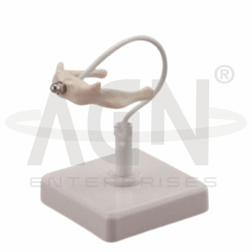
The Hyoid Bone Model from AGN Enterprises is an anatomical teaching aid designed to demonstrate the structure, shape, and anatomical features of the human hyoid bone. It helps students understand this distinctive U-shaped bone and its important relationships with the tongue, larynx, pharynx, and muscles of the neck.
The model is suitable for medical colleges, nursing institutes, dental colleges, physiotherapy colleges, paramedical institutions, anatomy laboratories, ENT education, and other health-science training centers.
The hyoid is a small U-shaped bone located in the anterior neck, generally between the mandible and larynx.
Unlike most bones in the human skeleton, the hyoid does not directly articulate with another bone. Instead, it is suspended and stabilized by muscles and ligaments.
The major anatomical parts of the hyoid bone include:
Body
Greater Horns (Greater Cornua)
Lesser Horns (Lesser Cornua)
These structures provide attachment sites for several muscles and ligaments associated with the tongue, pharynx, and larynx.
The body forms the central portion of the hyoid bone.
It provides attachment for several muscles associated with movements of the tongue, floor of the mouth, and laryngeal region.
The anatomical model helps learners identify the body and understand its relationship with the projecting horns.
The greater horns, also known as the greater cornua, extend posteriorly from the body on both sides.
They provide important attachment points for muscles and ligaments of the neck and pharyngeal region.
The model enables students to examine their shape, orientation, and relationship with the central body of the hyoid.
The lesser horns, or lesser cornua, are smaller projections located near the junction between the body and greater horns.
These structures serve as attachment points for ligaments and other soft-tissue structures.
A three-dimensional model makes the difference between the greater and lesser horns easier to identify.
The hyoid bone has important anatomical relationships with both the tongue and larynx.
Muscles attaching to the hyoid participate in movements involved in swallowing, speech, and positioning of the larynx and tongue.
Because of these relationships, the model is particularly useful when studying head and neck anatomy, oral anatomy, ENT anatomy, and speech-related structures.
Several muscles attach directly or indirectly to the hyoid bone. These include groups commonly classified as suprahyoid and infrahyoid muscles.
The suprahyoid muscles generally connect the hyoid with structures superior to it, while the infrahyoid muscles connect it with structures below.
Understanding these attachments helps students appreciate the functional role of the hyoid during swallowing and movements of the larynx.
The hyoid bone performs several important mechanical and supportive functions. It:
The Hyoid Bone Model provides students with a three-dimensional reference for studying a relatively small bone that can be difficult to understand from illustrations alone.
It can be used for lessons involving osteology, head and neck anatomy, laryngeal anatomy, muscular attachments, swallowing, speech, and ENT anatomy.
The model supports anatomical identification, practical demonstrations, spatial understanding, and examination preparation.
AGN Enterprises supplies anatomical models, skeletal teaching aids, pathological models, biological teaching materials, and educational laboratory equipment for medical colleges, nursing institutes, dental colleges, physiotherapy programs, and other training institutions.
Our anatomical teaching models provide clear three-dimensional representations for practical and classroom-based education.









