
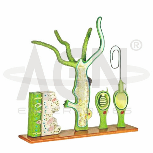
This 4-part model provides a detailed representation of Hydra anatomy through both longitudinal and cross-sectional views. It clearly highlights key structures such as the ectoderm, mesoglea, coelenteron, male and female gametes, buds, and mouth opening.
The cross-sectional block model, enlarged approximately 250X, illustrates various cellular components including nematoblasts, epithelial cells, sensory cells, interstitial cells, and the nerve network. An additional highly enlarged block model demonstrates the detailed structure of a cnidoblast cell.
Mounted on a board, this model is an excellent educational tool for studying cnidarian anatomy and cellular organization.









