
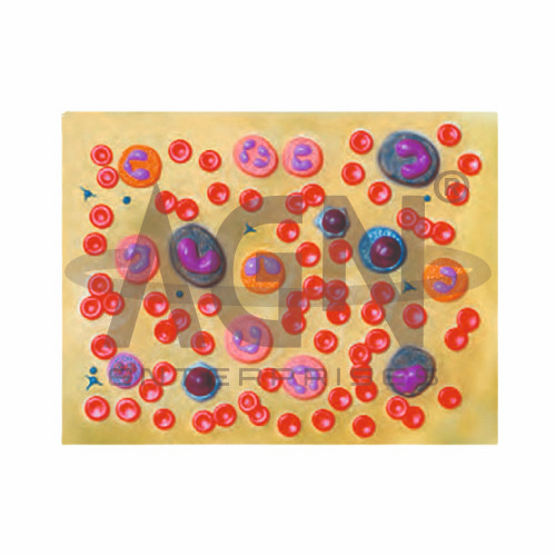
The Human Blood Cell Model from AGN Enterprises is an enlarged anatomical and biological teaching aid designed to demonstrate the major cellular components of human blood. The model helps students identify and compare red blood cells, different types of white blood cells, and platelets through clear three-dimensional representations.
It is suitable for medical colleges, nursing institutes, paramedical institutions, schools, biology laboratories, hematology departments, physiology laboratories, and health-science training centers.
Human blood is a specialized connective tissue consisting of plasma and formed elements. The formed elements include red blood cells, white blood cells, and platelets.
Depending on the specific model configuration, structures represented may include:
Red Blood Cells (Erythrocytes)
Neutrophils
Eosinophils
Basophils
Lymphocytes
Monocytes
Platelets (Thrombocytes)
The exact cells, colors, magnification, and structural details may vary according to the supplied model.
Red blood cells, or erythrocytes, are the most abundant formed elements in human blood. Mature human erythrocytes have a characteristic biconcave disc shape and normally lack a nucleus.
Their primary function is the transport of oxygen through hemoglobin. They also contribute to carbon dioxide transport.
The enlarged model helps students recognize the characteristic morphology of erythrocytes.
White blood cells, or leukocytes, are important components of the body’s immune and defense systems.
The major types of leukocytes include:
Neutrophils
Eosinophils
Basophils
Lymphocytes
Monocytes
An enlarged teaching model helps students compare their characteristic nuclear shapes, relative sizes, and cytoplasmic features.
Neutrophils, eosinophils, and basophils are collectively classified as granulocytes because characteristic granules are present in their cytoplasm.
Neutrophils are important in responses to many bacterial infections.
Eosinophils participate in immune responses involving parasites and allergic processes.
Basophils contain granules with substances involved in inflammatory and allergic responses.
The model helps learners distinguish these cell types visually.
Lymphocytes and monocytes are commonly grouped as agranulocytes based on their appearance under conventional light microscopy.
Lymphocytes play major roles in adaptive and other immune responses, while monocytes can migrate into tissues and differentiate into macrophages and related cells.
The model provides a useful visual reference for comparing these cells with granulocytes.
Platelets, or thrombocytes, are small cell fragments involved in hemostasis and blood clot formation.
They help form the initial platelet plug following blood-vessel injury and participate in processes that limit blood loss.
Where represented, the model allows students to compare their relatively small size with red and white blood cells.
The Human Blood Cell Model provides a three-dimensional approach to studying hematology and blood-cell morphology.
Teachers can use it for lessons involving blood composition, erythrocytes, leukocytes, platelets, immune function, hematology, physiology, and human biology.
The enlarged representation supports cell identification, comparative study, practical demonstrations, and examination preparation.
AGN Enterprises supplies anatomical models, biological teaching aids, cell models, pathological models, scientific instruments, and educational laboratory equipment for schools, medical colleges, nursing institutes, paramedical institutions, and training centers.
Our teaching models provide clear three-dimensional representations to support practical biology, anatomy, and medical education.









