
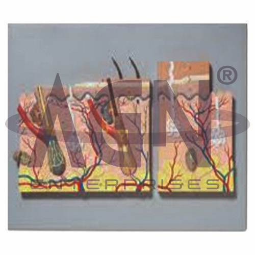
The Hairy & Hairless Skin Model from AGN Enterprises is an enlarged anatomical teaching aid designed to demonstrate and compare the microscopic organization of hairy skin and hairless (glabrous) skin. The model helps students understand the layers of the skin, hair follicles, glands, sensory structures, blood vessels, and other important components of the integumentary system.
It is suitable for medical colleges, nursing institutes, dermatology departments, paramedical institutions, cosmetology programs, biology laboratories, anatomy departments, and health-science training centers.
The Hairy & Hairless Skin Model provides a three-dimensional comparison of two important types of human skin.
Depending on the specific model configuration, represented structures may include:
Epidermis
Dermis
Subcutaneous Tissue
Hair Shaft
Hair Follicle
Hair Root
Sebaceous Gland
Sweat Glands
Arrector Pili Muscle
Blood Vessels
Sensory Nerves and Receptors
The exact structures, magnification, colors, and anatomical details may vary according to the supplied model.
The epidermis forms the outermost layer of the skin and consists primarily of keratinized stratified squamous epithelium.
Its layers vary in prominence according to the type and location of the skin. Hairless thick skin, such as that found on the palms and soles, has a particularly well-developed epidermis.
The enlarged model helps students visualize the relationship between the epidermis and deeper skin layers.
The dermis lies beneath the epidermis and consists mainly of connective tissue.
It provides structural support and contains numerous important structures, including blood vessels, nerves, sensory receptors, sweat glands, and hair-associated structures in hairy skin.
The model provides a clear representation of how these components are organized within the skin.
Hairy skin contains hair follicles and associated structures.
A typical hair follicle extends from the epidermal surface into deeper layers and is commonly associated with a sebaceous gland and arrector pili muscle.
The model helps learners understand the position and relationship of these structures in three dimensions.
Hairless or glabrous skin lacks hair follicles and is found in specialized areas such as the palms of the hands and soles of the feet.
These regions are adapted for functions involving friction, grip, and sensory perception. Thick glabrous skin has a relatively prominent epidermis and contains numerous sensory structures.
The comparative model makes differences between hairy and hairless skin easier to identify.
The model may demonstrate important glands associated with the skin.
Sweat glands contribute to thermoregulation and other skin functions.
Sebaceous glands, typically associated with hair follicles, produce sebum that helps lubricate the skin and hair.
Their enlarged representation helps students understand their location within the integumentary system.
Skin contains numerous sensory nerve endings and receptors that respond to stimuli such as touch, pressure, temperature, and pain.
Blood vessels within deeper skin layers provide nutrients and participate in temperature regulation.
Where represented, these structures help students connect skin anatomy with sensory and physiological functions.
The model is particularly useful for comparative study:
Hairy Skin – contains hair follicles and associated sebaceous glands and arrector pili muscles.
Hairless Skin – lacks hair follicles and is specialized for areas such as the palms and soles.
This side-by-side comparison helps learners recognize structural differences between major skin types.
The Hairy & Hairless Skin Model provides students with an enlarged three-dimensional approach to studying skin anatomy.
Teachers can use it for lessons involving epidermis, dermis, hair follicles, glands, sensory receptors, skin histology, dermatology, and the integumentary system.
The model supports anatomical identification, comparative study, practical demonstrations, and examination preparation.
AGN Enterprises supplies anatomical models, pathological models, biological teaching aids, scientific instruments, and educational laboratory equipment for medical colleges, nursing institutes, paramedical institutions, schools, and professional training centers.
Our teaching models provide clear three-dimensional representations for practical anatomy, biology, and medical education.









