
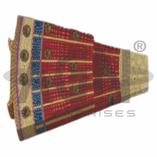
The Gymnosperm Stem (Pinus Silvestris) Model from AGN Enterprises is an enlarged botanical teaching aid designed to demonstrate the internal structure and tissue organization of a pine stem. The model helps students understand important anatomical features of a gymnosperm stem, including vascular tissues, secondary growth, annual rings, medullary rays, and resin canals.
It is suitable for schools, colleges, universities, botany laboratories, biology departments, forestry institutes, agricultural institutions, and educational science laboratories.
The Gymnosperm Stem (Pinus Silvestris) Model provides a three-dimensional representation of important tissues found in a typical pine stem.
Depending on the specific model configuration, structures represented may include:
Epidermis / Outer Protective Tissue
Cortex
Phloem
Vascular Cambium
Secondary Xylem
Annual Growth Rings
Medullary Rays
Resin Canals
Pith
The exact structures, colors, magnification, and sectional details may vary according to the supplied model.
In younger stems, the outer region includes epidermal and cortical tissues. As secondary growth progresses, protective tissues develop around the increasing stem circumference.
The enlarged model helps students understand how the outer tissues relate spatially to the vascular and central regions of the stem.
The cortex lies toward the outer portion of the stem and contains tissues involved in storage and other physiological functions.
The model provides a useful visual reference for identifying the cortex and distinguishing it from the vascular tissues located farther inward.
The vascular cambium is a lateral meristem responsible for secondary growth.
It produces secondary xylem toward the inside and secondary phloem toward the outside. Continued cambial activity contributes to an increase in stem diameter.
This makes the model particularly useful for teaching secondary growth in woody gymnosperms.
The secondary xylem forms the major woody portion of a mature pine stem.
In gymnosperms such as Pinus, water-conducting and supporting functions are performed primarily by tracheids rather than the vessel elements characteristic of many angiosperms.
The model helps students relate secondary xylem development to wood formation.
Successive periods of secondary xylem production can produce visible annual growth rings under seasonal growing conditions.
Differences between early-season and later-season wood contribute to the appearance of these rings.
Where represented, the model provides a clear demonstration of annual-ring formation and secondary growth.
Medullary or vascular rays extend radially through portions of the stem.
These tissues contribute to radial transport and storage within woody stems.
Their orientation makes them particularly useful for explaining the difference between longitudinal and radial tissue organization.
A characteristic anatomical feature of many Pinus species is the presence of resin canals or resin ducts.
These canals contain resin and contribute to the plant’s protective responses.
Where represented, the enlarged model makes their location within the stem easier for students to identify.
The pith occupies the central region of a young stem and consists largely of parenchymatous tissue.
Its position provides an important reference point when studying the concentric arrangement of vascular and other tissues in transverse section.
The Gymnosperm Stem (Pinus Silvestris) Model is particularly useful for explaining secondary growth.
A simplified sequence is:
Cambial Activity → Secondary Xylem Formation → Secondary Phloem Formation → Increase in Stem Diameter → Development of Woody Tissue
This helps students connect meristematic activity with the development of a woody stem.
The model provides an enlarged three-dimensional approach to studying gymnosperm stem anatomy.
Teachers can use it for lessons involving Pinus anatomy, secondary growth, vascular cambium, xylem, phloem, annual rings, medullary rays, resin canals, and gymnosperm botany.
It supports tissue identification, practical demonstrations, comparative plant anatomy, and examination preparation.
AGN Enterprises supplies botanical models, biological teaching aids, anatomical models, scientific instruments, and educational laboratory equipment for schools, colleges, universities, agricultural institutes, forestry programs, and professional laboratories.
Our botanical teaching models provide clear three-dimensional representations to support practical biology and plant anatomy education.









