
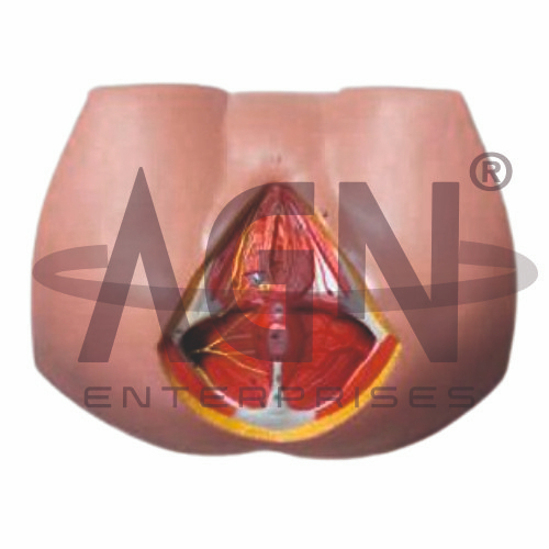
The Female Perineum Model from AGN Enterprises is a detailed anatomical teaching aid designed to demonstrate the muscles, fascia, external genital structures, pelvic floor, and associated anatomy of the female perineal region. The model helps students understand the organization and relationships of structures within this clinically important area.
It is suitable for medical colleges, nursing institutes, physiotherapy colleges, obstetrics and gynecology departments, paramedical institutions, anatomy laboratories, and health-science training centers.
The Female Perineum Model provides a three-dimensional representation of important structures associated with the female pelvic floor and external genital region.
Depending on the specific model configuration, structures represented may include:
Pelvic Floor Muscles
Perineal Muscles
External Genital Structures
Urethral Region
Vaginal Opening
Anal Region
Perineal Body
Associated Fascia
Blood Vessels and Nerves
The exact anatomical structures, colors, removable components, and level of detail may vary according to the supplied model.
The perineum is the anatomical region located inferior to the pelvic diaphragm between the thighs.
For anatomical study, it is commonly divided into two regions:
Urogenital Triangle
Anal Triangle
The model helps students visualize these divisions and understand the positions of important structures within each region.
The urogenital triangle forms the anterior portion of the female perineum.
It contains structures associated with the external genitalia and openings of the urinary and reproductive tracts.
The model provides a useful three-dimensional reference for studying these structures and their relationships with surrounding muscles and soft tissues.
The pelvic floor consists of muscles and connective tissues that provide important support to the pelvic organs.
The levator ani muscle group forms a major component of the pelvic diaphragm, together with associated muscles and fascia.
Where represented, the model helps learners understand how these structures contribute to pelvic support and function.
Several muscles contribute to the structure and function of the female perineum.
Depending on the model, represented muscles may include structures associated with the superficial and deep perineal regions.
These muscles contribute to pelvic support and functions associated with the urinary, reproductive, and anorectal systems.
The perineal body is a central fibromuscular structure located between the vaginal opening and anus.
Several muscles and fascial structures attach to this region, making it important for maintaining pelvic-floor integrity.
The model can help students understand its position and significance in female pelvic anatomy.
The Female Perineum Model is particularly useful for teaching clinical anatomy related to obstetrics, gynecology, pelvic-floor rehabilitation, and women’s health physiotherapy.
It can support explanations of perineal anatomy during childbirth, pelvic-floor function, and the relationships between reproductive, urinary, and anorectal structures.
The model is intended for anatomical education rather than diagnosis.
The Female Perineum Model provides students with a three-dimensional approach to studying an anatomically complex region.
Teachers can use it for lessons involving female perineal anatomy, pelvic floor, perineal muscles, external genital anatomy, urogenital triangle, anal triangle, obstetric anatomy, and gynecology.
The model supports anatomical identification, spatial visualization, practical demonstrations, and examination preparation.
AGN Enterprises supplies anatomical models, reproductive-system models, pelvic models, biological teaching aids, and educational laboratory equipment for medical colleges, nursing institutes, physiotherapy programs, paramedical institutions, and professional training centers.
Our anatomical models provide clear three-dimensional representations for practical and classroom-based medical education.









