
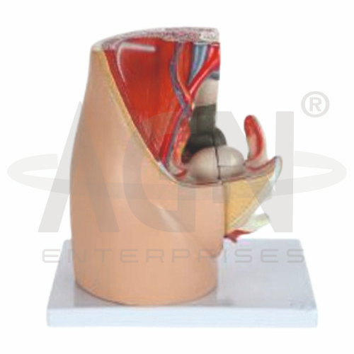
The Female Pelvis Model from AGN Enterprises is a detailed anatomical teaching aid designed to demonstrate the bones, joints, pelvic cavity, and important structural features of the female pelvis. The model provides a three-dimensional representation that helps students understand female pelvic anatomy and its relationship with the spine, hip joints, pelvic floor, and reproductive region.
It is suitable for medical colleges, nursing institutes, physiotherapy colleges, obstetrics and gynecology departments, paramedical institutions, anatomy laboratories, and health-science training centers.
The Female Pelvis Model provides a clear representation of the major bones and landmarks forming the female pelvic girdle.
Depending on the specific model configuration, structures represented may include:
Ilium
Ischium
Pubis
Sacrum
Coccyx
Pubic Symphysis
Sacroiliac Joints
Acetabulum
Pelvic Inlet
Pelvic Outlet
The exact structures, articulation, mounting, and anatomical details may vary according to the supplied model.
The pelvis is formed by the paired hip bones together with the sacrum and coccyx.
Each hip bone develops from three principal components:
Ilium
Ischium
Pubis
These components unite around the acetabulum, which articulates with the head of the femur to form the hip joint.
The pelvic inlet forms the superior opening of the true pelvis.
Its boundaries and dimensions are particularly important in obstetric anatomy because they contribute to the bony passage associated with childbirth.
The model allows students to visualize the shape and orientation of the pelvic inlet in three dimensions.
The pelvic outlet forms the inferior opening of the true pelvis.
Studying the relationship between the pelvic inlet, pelvic cavity, and pelvic outlet provides an important foundation for understanding female pelvic and obstetric anatomy.
The model offers a practical visual reference for these anatomical regions.
The sacrum forms the posterior portion of the pelvis and connects the vertebral column with the pelvic girdle.
The coccyx lies inferior to the sacrum and forms the terminal portion of the vertebral column.
These structures contribute to the posterior boundaries and stability of the pelvis.
The pubic symphysis is a cartilaginous joint connecting the right and left pubic bones anteriorly.
The model helps students understand its location and relationship with the pelvic cavity and surrounding bony landmarks.
This structure is particularly relevant in musculoskeletal and obstetric anatomy.
The sacrum articulates with the ilium on each side at the sacroiliac joints.
These joints transfer loads between the vertebral column and lower limbs and contribute to pelvic stability.
Their position can be studied clearly using a three-dimensional female pelvis model.
The Female Pelvis Model is particularly useful for teaching the bony anatomy relevant to obstetrics.
Teachers can use it to explain concepts involving the pelvic inlet, pelvic cavity, pelvic outlet, sacrum, pubic symphysis, and general birth-canal dimensions.
It provides a useful foundation for nursing, midwifery, obstetrics, and gynecology education.
The model provides students with a three-dimensional approach to studying female pelvic anatomy.
Teachers can use it for lessons involving pelvic bones, pelvic joints, pelvic inlet and outlet, hip anatomy, obstetric anatomy, musculoskeletal anatomy, physiotherapy, and gynecology.
The model supports anatomical identification, spatial visualization, practical demonstrations, and examination preparation.
AGN Enterprises supplies anatomical models, pelvic models, skeletal teaching aids, reproductive-system models, joint models, and educational laboratory equipment for medical colleges, nursing institutes, physiotherapy programs, paramedical institutions, and professional training centers.
Our anatomical teaching models provide clear three-dimensional representations for practical and classroom-based medical education.









