
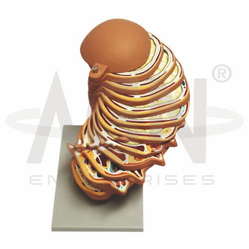
The Disc Head Model from AGN Enterprises is an anatomical teaching aid designed to demonstrate the sectional anatomy and important internal structures of the human head. Its disc or sectional presentation helps students understand the spatial relationships between the brain, skull, facial structures, cavities, muscles, and other anatomical components of the head.
The model is suitable for medical colleges, dental colleges, nursing institutes, physiotherapy colleges, paramedical institutions, anatomy laboratories, biology departments, and health-science training centers.
The Disc Head Model provides a three-dimensional representation of selected structures of the head in sectional form.
Depending on the specific model configuration, represented structures may include:
Brain
Cranial Cavity
Skull Structures
Nasal Cavity
Oral Cavity
Pharyngeal Region
Facial Muscles
Head and Neck Muscles
Blood Vessels
Nerves
The exact sectional arrangement, anatomical structures, colors, removable components, and level of detail may vary according to the supplied model.
The sectional design helps learners visualize structures located at different depths within the head.
Unlike a completely external anatomical model, a disc-head representation can provide access to deeper structures and demonstrate how the brain, skull, facial cavities, muscles, nerves, and vessels relate spatially.
This makes it particularly useful for practical anatomy demonstrations.
Where represented, the model demonstrates portions of the brain within the cranial cavity.
Students can examine the relationship between neural structures and the surrounding skull while gaining a better understanding of their anatomical orientation.
The model can complement separate brain and skull models during neuroanatomy lessons.
The nasal and oral cavities occupy important regions of the facial skeleton.
A sectional head model helps students visualize their relative positions and relationships with the palate, pharynx, and surrounding structures.
This is useful for studying respiratory, digestive, dental, and head-and-neck anatomy.
Depending on the configuration, the model may demonstrate important muscles associated with the face, mastication, scalp, and neck.
These structures help learners understand the muscular organization responsible for facial expression, chewing, jaw movement, and stabilization of the head.
Where represented, nerves and blood vessels provide additional detail for studying the neurovascular anatomy of the head.
Their inclusion helps students understand how neural and vascular structures relate to muscles, bones, cavities, and other tissues.
This can be particularly useful for medical and dental anatomy education.
The Disc Head Model can help students understand the transition between structures of the head and upper neck.
It provides useful context for studying relationships involving the pharynx, oral cavity, cranial structures, muscles, nerves, and associated anatomical regions.
The model provides students with a three-dimensional approach to studying complex sectional head anatomy.
Teachers can use it for lessons involving brain anatomy, cranial anatomy, facial structures, nasal and oral cavities, head musculature, nerves, blood vessels, and head-and-neck anatomy.
It supports anatomical identification, spatial visualization, practical demonstrations, and examination preparation.
AGN Enterprises supplies anatomical models, head-and-neck models, skull models, neurological models, muscular teaching aids, and educational laboratory equipment for medical colleges, dental colleges, nursing institutes, physiotherapy programs, and professional training centers.
Our anatomical teaching models provide clear three-dimensional representations for practical and classroom-based human anatomy education.









