
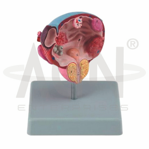
The Diseases of the Urinary Bladder & Prostate Model from AGN Enterprises is a detailed pathological teaching aid designed to demonstrate common diseases and structural changes affecting the urinary bladder and prostate gland. The model helps students compare normal anatomy with selected pathological conditions and understand the close anatomical relationship between the prostate, urethra, and urinary bladder.
It is suitable for medical colleges, nursing institutes, paramedical institutions, urology departments, pathology laboratories, anatomy departments, and health-science training centers.
The model provides a three-dimensional representation of selected normal and pathological structures involving the urinary bladder and prostate gland.
Depending on the specific model configuration, represented conditions and structures may include:
Urinary Bladder
Prostate Gland
Prostatic Urethra
Benign Prostatic Hyperplasia (BPH)
Prostate Tumor Changes
Bladder Stones
Bladder Tumor Changes
Inflammatory or Other Pathological Features
The exact diseases, stages, colors, sections, and pathological details may vary according to the supplied model.
The urinary bladder is a hollow muscular organ that temporarily stores urine before urination.
Urine reaches the bladder through the ureters and leaves through the urethra.
The model helps learners understand the position of the bladder and its relationship with the prostate and lower urinary tract.
The prostate is a gland of the male reproductive system located inferior to the urinary bladder.
The proximal urethra passes through the prostate. Because of this anatomical relationship, enlargement of the prostate can affect the passage of urine.
The model provides a useful three-dimensional reference for studying this clinically important region.
Benign prostatic hyperplasia (BPH) is a noncancerous enlargement of the prostate that becomes increasingly common with age.
Prostatic enlargement can narrow or compress the urethral passage and contribute to lower urinary tract symptoms.
Where represented, the model helps students compare an enlarged prostate with normal anatomy.
Prostate cancer is a malignant disease arising from cells of the prostate gland.
Where pathological changes are represented, the model can support introductory teaching about the differences between benign enlargement and malignant prostate disease.
The model is intended for anatomical and pathological education rather than diagnosis.
Bladder stones are hardened mineral deposits that can form within the urinary bladder.
They may develop when the bladder does not empty completely or under other clinical circumstances.
Where represented, the model provides a visual reference for explaining their location within the bladder.
Tumors can develop from tissues of the urinary bladder, with many bladder cancers arising from the urothelial lining.
Where represented, pathological sections can help students understand how abnormal growth may alter the internal structure of the bladder.
One of the main educational benefits of the Diseases of the Urinary Bladder & Prostate Model is its demonstration of the close relationship between these organs.
Students can visualize how changes in the prostate may influence the urethra and urinary outflow from the bladder, providing useful context for urology and pathology education.
The model provides students with a three-dimensional approach to studying urinary bladder and prostate pathology.
Teachers can use it for lessons involving bladder anatomy, prostate anatomy, BPH, prostate disease, bladder stones, bladder tumors, urinary obstruction, pathology, and urology.
It supports pathological identification, comparison with normal anatomy, clinical demonstrations, and examination preparation.
AGN Enterprises supplies pathological models, anatomical models, urinary-system models, reproductive-system models, scientific instruments, and educational laboratory equipment for medical colleges, nursing institutes, paramedical institutions, hospitals, and professional training centers.
Our teaching models provide clear three-dimensional representations to support practical anatomy, pathology, and medical education.









