
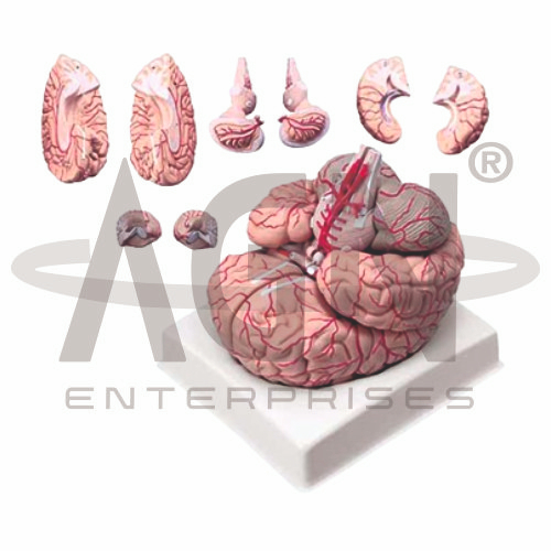
The Brain with Arteries 9 Parts Model from AGN Enterprises is a detailed multi-part neuroanatomical teaching aid designed to demonstrate the major structures of the human brain together with its arterial blood supply. Its nine-part construction allows selected brain components to be separated for closer examination of external, internal, and vascular anatomy.
The model is suitable for medical colleges, nursing institutes, physiotherapy colleges, neuroscience departments, neurology programs, anatomy laboratories, paramedical institutions, and health-science training centers.
The Brain with Arteries 9 Parts Model provides a three-dimensional representation of the brain and its major arterial structures.
Depending on the specific model configuration, represented structures may include:
Cerebral Hemispheres
Frontal Lobe
Parietal Lobe
Temporal Lobe
Occipital Lobe
Cerebellum
Brainstem
Major Cerebral Arteries
Circle of Willis Region
The exact division of the nine parts, arterial branches, colors, removable components, and anatomical details may vary according to the supplied model.
A major feature of the model is its nine-part separable design.
The multiple components provide improved access to structures that may be difficult to observe on a single-piece brain model. Students can remove selected sections, examine their anatomy, and reposition them to understand their normal spatial relationships.
This makes the model especially useful for practical neuroanatomy and cerebral circulation studies.
The cerebrum consists of right and left cerebral hemispheres, which form the largest portion of the human brain.
Their surfaces contain numerous gyri and sulci that increase the surface area of the cerebral cortex.
The model helps students examine the external morphology of the hemispheres alongside their arterial supply.
The model can support identification of the major cerebral lobes:
Frontal Lobe
Parietal Lobe
Temporal Lobe
Occipital Lobe
These regions participate in numerous functions involving voluntary movement, sensation, cognition, language, memory, hearing, and vision.
The cerebellum is located posterior to the brainstem and inferior to the posterior cerebrum.
It contributes to motor coordination, balance, posture, timing of movement, and motor learning.
Where removable, the cerebellar component can be examined separately to better understand its relationship with the brainstem and cerebral hemispheres.
The brainstem connects the brain with the spinal cord and includes the midbrain, pons, and medulla oblongata.
It contains important neural pathways and centers associated with sensory, motor, cranial nerve, respiratory, and cardiovascular functions.
The model helps learners understand its position relative to the cerebrum and cerebellum.
The brain receives its arterial blood supply primarily through the internal carotid and vertebrobasilar circulations.
Major arteries represented on the model may include:
Anterior Cerebral Arteries
Middle Cerebral Arteries
Posterior Cerebral Arteries
Vertebral Arteries
Basilar Artery
Communicating Arteries
The exact vessels displayed depend on the supplied model.
Where represented, the Circle of Willis can be studied at the base of the brain as an important arterial network.
It connects components of the anterior and posterior cerebral circulations through communicating arteries.
The model provides a useful three-dimensional reference for understanding this complex vascular arrangement.
The anterior cerebral artery supplies important medial regions of the frontal and parietal lobes.
Its position and relationship with other arteries can be studied more effectively when vascular anatomy is displayed directly on a three-dimensional brain model.
The middle cerebral artery supplies extensive portions of the lateral cerebral hemisphere.
Its branches are particularly important in clinical neuroanatomy and cerebrovascular education.
The model helps students follow its course in relation to major cerebral regions.
The posterior cerebral artery contributes to the blood supply of posterior and inferior cerebral regions, including important areas of the occipital and temporal lobes.
The model allows students to compare posterior cerebral circulation with vessels arising from the internal carotid circulation.
The vertebral arteries contribute to the basilar artery, forming an important component of the posterior circulation.
This vascular system supplies regions associated with the brainstem, cerebellum, and posterior portions of the brain.
Where represented, the model provides a clear spatial reference for these vessels.
The Brain with Arteries 9 Parts Model can provide an anatomical foundation for teaching cerebral circulation, arterial territories, stroke anatomy, vascular lesions, brainstem circulation, and other cerebrovascular topics.
Its multi-part construction allows students to relate individual brain regions to the vessels supplying them.
The model is intended for educational demonstration rather than diagnosis.
The model provides a comprehensive three-dimensional approach to studying both structural and vascular neuroanatomy.
Teachers can use it for lessons involving brain lobes, cerebral hemispheres, cerebellum, brainstem, cerebral arteries, Circle of Willis, carotid circulation, and vertebrobasilar circulation.
Its nine-part design supports hands-on learning, anatomical identification, spatial visualization, practical demonstrations, and examination preparation.
AGN Enterprises supplies brain models, neurological models, vascular models, nervous-system teaching aids, anatomical models, scientific instruments, and educational laboratory equipment for medical colleges, nursing institutes, universities, and professional training centers.
Our anatomical models provide clear three-dimensional representations for practical neuroanatomy and medical education.









