
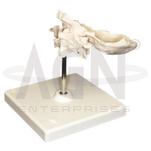
The Axis and Occipital Bone (Atlas) Model from AGN Enterprises is a detailed anatomical teaching aid designed to demonstrate the occipital bone, atlas (C1), axis (C2), and their important articulations in the upper cervical spine. The model helps students understand the anatomical relationships responsible for supporting the skull and permitting movements of the head and neck.
It is suitable for medical colleges, nursing institutes, physiotherapy colleges, orthopedic departments, chiropractic education, paramedical institutions, anatomy laboratories, and health-science training centers.
The model provides a three-dimensional representation of the craniovertebral junction, where the skull connects with the cervical vertebral column.
Depending on the specific model configuration, represented structures may include:
Occipital Bone
Occipital Condyles
Atlas (C1)
Axis (C2)
Dens or Odontoid Process
Atlanto-Occipital Joint
Atlanto-Axial Joint
Vertebral Foramina
Transverse Processes
Associated Articular Surfaces
The exact structures, ligaments, movable components, colors, and anatomical details may vary according to the supplied model.
The occipital bone forms much of the posterior and inferior portion of the skull.
Its inferior surface contains the occipital condyles, which articulate with the superior articular surfaces of the atlas.
This relationship forms the paired atlanto-occipital joints and connects the skull to the vertebral column.
The atlas (C1) is the first cervical vertebra and directly supports the skull.
Unlike a typical vertebra, the atlas does not have a conventional vertebral body or prominent spinous process. It consists mainly of anterior and posterior arches connected by lateral masses.
Its superior articular surfaces articulate with the occipital condyles.
The axis (C2) is the second cervical vertebra and forms an important pivot for rotation of the head and atlas.
Its most distinctive anatomical feature is the dens, also called the odontoid process.
The model provides a clear three-dimensional reference for studying the relationship between the dens and atlas.
The dens projects superiorly from the body of the axis.
It acts as a pivot around which the atlas and skull can rotate, making it an important structure in upper cervical biomechanics.
Where movement is incorporated into the model, this relationship can be demonstrated practically.
The atlanto-occipital joints are formed between the occipital condyles of the skull and the superior articular surfaces of the atlas.
These joints primarily permit flexion and extension of the head, including the movement commonly associated with nodding “yes.”
They also allow a limited degree of lateral flexion.
The atlanto-axial articulation is formed between the atlas and axis.
The median atlanto-axial joint involves the dens and the anterior arch of the atlas, while paired lateral articulations occur between the articular surfaces of C1 and C2.
This joint complex permits substantial rotation of the head and atlas relative to the axis.
The model can be used to explain important movements occurring at the craniovertebral junction, including:
The atlanto-occipital region contributes prominently to nodding movements, while the atlanto-axial region contributes substantially to head rotation.
The atlas and axis differ considerably from the remaining cervical vertebrae because of their specialized roles in supporting the skull and providing mobility.
Studying these structures together with the occipital bone helps learners understand the transition between the skull and vertebral column.
The model can provide an anatomical reference when teaching topics involving upper cervical biomechanics, atlas and axis injuries, odontoid fractures, craniovertebral stability, and cervical-spine assessment.
It is particularly useful for orthopedic, physiotherapy, and musculoskeletal anatomy education.
The model is intended for educational demonstration rather than diagnosis.
The Axis and Occipital Bone (Atlas) Model provides students with a three-dimensional approach to studying the craniovertebral junction.
Teachers can use it for lessons involving occipital bone, atlas, axis, dens, atlanto-occipital joint, atlanto-axial joint, cervical vertebrae, and head and neck movements.
It supports anatomical identification, spatial visualization, biomechanics demonstrations, and examination preparation.
AGN Enterprises supplies spinal models, skull models, skeletal teaching aids, joint models, anatomical models, scientific instruments, and educational laboratory equipment for medical colleges, nursing institutes, physiotherapy programs, universities, and professional training centers.
Our anatomical teaching models provide clear three-dimensional representations for practical musculoskeletal and human anatomy education.









