
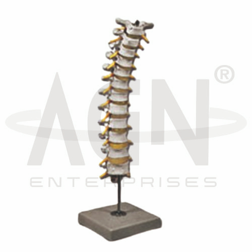
The Thoracic Spinal Column Model from AGN Enterprises is a detailed anatomical teaching aid designed to demonstrate the thoracic region of the human vertebral column. It helps students understand the structure and arrangement of the thoracic vertebrae, intervertebral spaces, vertebral canal, and associated spinal structures.
The model is suitable for medical colleges, nursing institutes, physiotherapy colleges, paramedical institutions, anatomy laboratories, orthopedic education, biology departments, and health-science training centers.
The thoracic spine forms the middle region of the vertebral column and normally consists of 12 thoracic vertebrae, designated T1 to T12.
Depending on the specific model configuration, structures represented may include:
Thoracic Vertebrae
Vertebral Bodies
Spinous Processes
Transverse Processes
Articular Processes
Intervertebral Discs
Vertebral Canal
Costal Facets
Associated Spinal Structures
The exact structures and level of anatomical detail may vary according to the supplied model.
Thoracic vertebrae are located between the cervical and lumbar regions of the vertebral column.
Each typical thoracic vertebra contains a vertebral body, vertebral arch, spinous process, transverse processes, and articular processes.
The three-dimensional model allows students to examine these structures and understand how successive vertebrae form the thoracic spinal column.
The thoracic spinal column consists of twelve vertebrae:
T1 – T12
These vertebrae gradually change in size and morphology from the upper to lower thoracic region.
Studying them as part of a continuous spinal column helps students understand the transition between the cervical, thoracic, and lumbar regions.
Where represented, intervertebral discs can be observed between adjacent vertebral bodies.
These fibrocartilaginous structures contribute to flexibility, distribute mechanical loads, and provide cushioning between vertebrae.
The model helps learners visualize the position of the discs in relation to the vertebral bodies.
The vertebral foramina of successive vertebrae collectively form the vertebral canal.
This canal provides protection for the spinal cord and associated structures.
The model can help students understand how the bony components of the spinal column form a protective passage for neural structures.
A characteristic feature of thoracic vertebrae is their anatomical relationship with the ribs.
Thoracic vertebrae possess costal facets that participate in articulations with the ribs. Together, the thoracic vertebrae, ribs, and sternum contribute to the framework of the thoracic cage.
This relationship makes the thoracic spine important in both vertebral and thoracic anatomy.
The thoracic spinal column performs several important functions, including:
Supporting the Upper Body
Protecting Neural Structures
Providing Attachment and Articulation for Ribs
Contributing to Posture
Allowing Controlled Spinal Movement
Helping Form the Posterior Thoracic Cage
The model allows these structural and functional concepts to be demonstrated in three dimensions.
The Thoracic Spinal Column Model provides students with a practical approach to studying the middle region of the vertebral column.
Teachers can use it for lessons involving vertebral anatomy, thoracic spine, intervertebral discs, rib articulations, vertebral canal, musculoskeletal anatomy, orthopedics, and physiotherapy.
The model supports anatomical identification, spatial visualization, practical demonstrations, and examination preparation.
AGN Enterprises supplies anatomical models, skeletal teaching aids, biological models, medical teaching equipment, and educational laboratory products for medical colleges, nursing institutes, physiotherapy programs, paramedical institutions, and training centers.
Our anatomical models provide clear three-dimensional representations for practical and classroom-based human anatomy education.









