
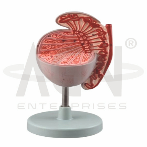
The Testis (3.5x Life Size) Model from AGN Enterprises is an enlarged anatomical teaching aid designed to demonstrate the internal and external structures of the human testis at approximately 3.5 times natural size. The enlarged format makes small reproductive structures easier to identify and understand during classroom and laboratory demonstrations.
The model is suitable for medical colleges, nursing institutes, paramedical institutions, biology laboratories, anatomy departments, urology education, reproductive biology programs, and health-science training centers.
The testes are the primary male reproductive organs responsible for sperm production and testosterone secretion.
Depending on the specific model configuration, structures represented may include:
Testicular Lobules
Seminiferous Tubules
Rete Testis
Epididymis
Vas Deferens
Tunica Albuginea
Associated Blood Vessels
Spermatic Structures
The exact anatomical details may vary according to the supplied model.
A major feature of the Testis (3.5x Life Size) Model is its enlarged construction.
At approximately 3.5 times natural size, the model allows students to examine anatomical structures that would be difficult to observe clearly on a life-size representation.
The enlarged format is particularly useful for group demonstrations and detailed reproductive anatomy education.
The internal structure of the testis consists of numerous lobules containing highly coiled seminiferous tubules.
These tubules are associated with sperm production. Developing sperm cells eventually pass through a system of ducts toward the epididymis.
Where sectional anatomy is represented, the model helps learners understand these internal pathways in three dimensions.
The epididymis is a highly coiled duct associated with the posterior aspect of the testis.
It plays an important role in the maturation, storage, and transport of sperm.
The enlarged model helps students visualize the relationship between the testis and the different regions of the epididymis.
The vas deferens continues from the epididymal duct and transports sperm toward the pelvic reproductive tract.
A simplified pathway can be demonstrated as:
Seminiferous Tubules → Rete Testis → Efferent Ductules → Epididymis → Vas Deferens
This helps students connect individual anatomical structures with the pathway followed by sperm.
The seminiferous tubules are microscopic structures within the testes where spermatogenesis takes place.
Although their actual microscopic organization cannot be represented at natural scale on a gross anatomical model, an enlarged teaching model may illustrate their general arrangement within the testicular lobules.
This makes the model useful for connecting gross anatomy with introductory reproductive histology.
The testes have two major reproductive functions:
Spermatogenesis – production of male reproductive cells.
Hormone Production – secretion of androgens, particularly testosterone.
The model helps teachers connect these physiological functions with the anatomical organization of the testis.
The Testis (3.5x Life Size) Model provides an enlarged three-dimensional approach to male reproductive anatomy.
Teachers can use it for lessons involving testicular anatomy, spermatogenesis, seminiferous tubules, epididymis, vas deferens, male reproductive physiology, urology, and human reproduction.
The enlarged format supports anatomical identification, spatial visualization, practical demonstrations, and examination preparation.
AGN Enterprises supplies anatomical models, reproductive-system models, biological teaching aids, pathological models, and educational laboratory equipment for medical colleges, nursing institutes, paramedical institutions, schools, and professional training centers.
Our enlarged anatomical models help students examine complex human-body structures through clear three-dimensional representations.









