
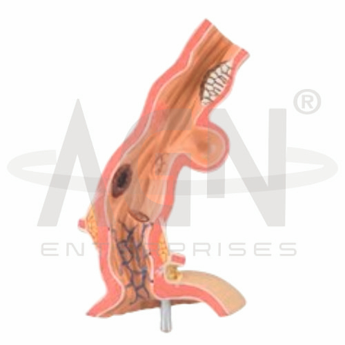
The Pathological Model of Esophagus from AGN Enterprises is a three-dimensional anatomical teaching model designed to demonstrate the structure of the human esophagus and representative pathological changes affecting the esophageal wall and lumen. The model helps students compare normal anatomy with disease-related abnormalities and understand their location and structural effects. It is suitable for medical colleges, nursing institutes, paramedical institutions, anatomy and pathology laboratories, gastroenterology education, and health-science training.
The Pathological Model of Esophagus provides a visual representation of esophageal anatomy along with selected pathological conditions.
Depending on the specific model configuration, it may demonstrate representative conditions such as:
Esophagitis
Esophageal Ulceration
Gastroesophageal Reflux-Related Changes
Barrett’s Esophagus
Esophageal Varices
Esophageal Carcinoma
Other Esophageal Abnormalities
The three-dimensional presentation makes pathological features easier to visualize and discuss during classroom demonstrations.
The esophagus is a muscular tube that carries swallowed food and liquids from the pharynx toward the stomach.
The model can help students study structures represented in the supplied configuration, including:
Esophageal Lumen
Mucosal Layer
Submucosal Layer
Muscular Layers
Outer Esophageal Wall
Connection toward the Stomach
Understanding normal esophageal anatomy provides an important foundation for studying pathological changes.
The Pathological Model of Esophagus can help teachers explain how different conditions affect the esophageal wall and passageway.
Inflammatory conditions can damage the esophageal lining, while chronic reflux can contribute to changes in the lower esophagus. Other conditions may produce abnormal dilation of veins, narrowing of the lumen, ulceration, or abnormal tissue growth.
Where represented on the supplied model, these abnormalities can be compared visually with normal esophageal structures.
The Pathological Model of Esophagus converts complex gastrointestinal pathology into a clear three-dimensional learning experience. Students can observe representative abnormalities and relate them to normal esophageal anatomy.
Teachers can use the model for lessons involving digestive-system anatomy, esophageal diseases, gastroenterology, pathology, oncology, and human anatomy.
The model is intended for educational demonstration purposes and is not designed for diagnosis or clinical decision-making.
AGN Enterprises supplies anatomical models, pathological models, biology teaching aids, and educational laboratory equipment for medical colleges, nursing institutes, paramedical institutions, schools, and professional training centers.
Our anatomical teaching models help learners connect theoretical medical concepts with clear three-dimensional representations.









