
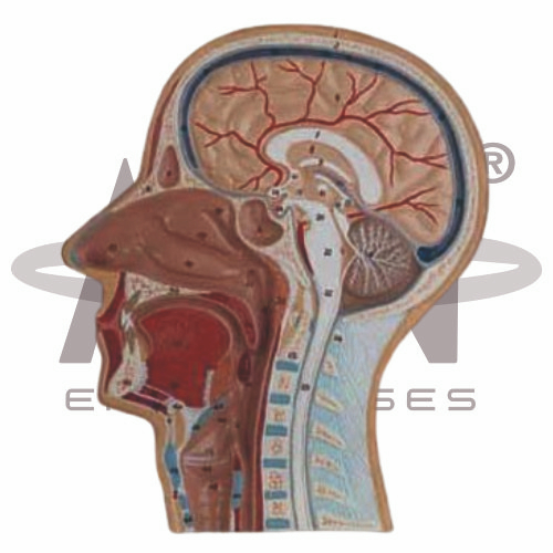
The Median Section of Head (Relief Model) from AGN Enterprises is a detailed anatomical teaching aid designed to demonstrate the internal structures of the human head and neck in a median or sagittal section. The relief-style presentation helps students visualize the spatial relationships between the brain, nasal cavity, oral cavity, pharynx, tongue, palate, vertebral region, and other structures of the head and upper neck.
The model is suitable for medical colleges, nursing institutes, paramedical institutions, biology laboratories, anatomy departments, ENT education, and other health-science training programs.
A median section divides the head along the midline and exposes important internal structures that cannot normally be viewed externally.
Depending on the specific model configuration, structures represented may include:
Brain
Nasal Cavity
Nasal Septum
Oral Cavity
Tongue
Hard and Soft Palate
Pharynx
Laryngeal Region
Cervical Vertebrae
Spinal Cord
The exact anatomical structures and level of detail may vary according to the supplied model.
The relief model can help students understand the position of the brain within the cranial cavity and its relationship with structures of the face and upper neck.
Where represented, learners may identify structures such as the cerebrum, cerebellum, brainstem, and associated cranial regions.
The sectional presentation is particularly useful for understanding how the brain connects inferiorly with the spinal cord.
The Median Section of Head (Relief Model) provides a useful visual representation of the relationship between the nasal and oral cavities.
Students can examine structures such as the nasal cavity, palate, tongue, and oral cavity and understand their positions relative to one another.
This makes the model valuable for lessons involving respiration, swallowing, speech, and head-and-neck anatomy.
The pharynx forms an important shared region associated with both the respiratory and digestive systems.
The model can help students visualize the relationship between the:
Nasal Cavity
Oral Cavity
Pharynx
Upper Airway
Esophageal Region
This sectional view makes complex internal pathways easier to understand than with external anatomy models alone.
One of the key benefits of this teaching aid is its ability to display several anatomical systems within a single sectional view.
Students can study how the nervous, respiratory, digestive, skeletal, and muscular structures of the head and neck relate spatially to one another.
The relief format provides depth while retaining a clear sectional presentation suitable for classroom demonstrations.
This anatomical model helps students move beyond two-dimensional textbook illustrations and develop a clearer understanding of median-sectional head anatomy.
Teachers can use it for lessons involving human anatomy, head-and-neck anatomy, neuroanatomy, nasal anatomy, oral anatomy, pharyngeal anatomy, ENT studies, and respiratory and digestive pathways.
It supports anatomical identification, spatial visualization, revision, and practical classroom demonstrations.
AGN Enterprises supplies anatomical models, biological teaching aids, pathological models, and educational laboratory equipment for medical colleges, nursing institutes, schools, paramedical institutions, and professional training centers.
Our anatomical teaching aids provide clear visual representations that help learners understand complex human-body structures and their relationships.









