
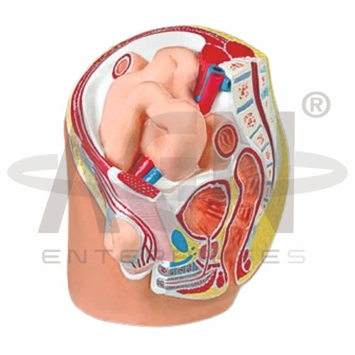
The Indirect Inguinal Hernia Model from AGN Enterprises is a specialized anatomical and pathological teaching aid designed to demonstrate the anatomical pathway and presentation of an indirect inguinal hernia. It helps students understand the relationship between the inguinal canal, deep inguinal ring, abdominal wall, and herniating abdominal contents.
The model is suitable for medical colleges, nursing institutes, paramedical institutions, surgical training centers, anatomy departments, pathology laboratories, and health-science education.
An indirect inguinal hernia occurs when abdominal contents protrude through the deep inguinal ring and travel along the inguinal canal.
Depending on the specific model configuration, structures represented may include:
Inguinal Canal
Deep Inguinal Ring
Superficial Inguinal Ring
Hernial Sac
Abdominal Wall
Spermatic Cord Region
Inguinal Ligament
Herniating Abdominal Contents
The exact anatomical and pathological structures represented may vary according to the supplied model.
The inguinal canal is an oblique passage through the lower anterior abdominal wall.
In males, it transmits the spermatic cord, while in females it contains the round ligament of the uterus along with associated structures.
The model helps learners visualize how an indirect hernia follows the course of this canal.
The Indirect Inguinal Hernia Model can be used to demonstrate the characteristic pathway of this type of hernia.
A simplified pathway is:
Abdominal Cavity → Deep Inguinal Ring → Inguinal Canal → Superficial Inguinal Ring
In some cases, the hernial sac may extend farther into the scrotal region in males.
An important anatomical feature used to distinguish inguinal hernias is their relationship to the inferior epigastric vessels.
An indirect inguinal hernia enters the deep inguinal ring lateral to the inferior epigastric vessels.
This anatomical relationship is particularly important in medical, surgical, and clinical anatomy education.
The model can help instructors explain the distinction between indirect and direct inguinal hernias.
Indirect Inguinal Hernia – enters through the deep inguinal ring and is lateral to the inferior epigastric vessels.
Direct Inguinal Hernia – protrudes through a weakened area of the posterior wall of the inguinal canal and is generally medial to the inferior epigastric vessels.
Understanding this difference is important in clinical and surgical anatomy.
The hernial sac is a peritoneal outpouching that accompanies the protruding contents.
Depending on the extent of the hernia, the sac may remain within the inguinal canal or extend through the superficial inguinal ring.
A three-dimensional model helps students visualize this pathway more effectively than diagrams alone.
The Indirect Inguinal Hernia Model provides a three-dimensional approach to studying an important clinical condition involving the abdominal wall and inguinal region.
Teachers can use it for lessons involving inguinal anatomy, hernia pathology, abdominal-wall anatomy, inguinal canal, clinical anatomy, and introductory surgical education.
The model supports anatomical identification, pathological demonstrations, clinical correlation, and examination preparation.
AGN Enterprises supplies anatomical models, pathological models, surgical teaching aids, biological models, and educational laboratory equipment for medical colleges, nursing institutes, paramedical institutions, and professional training centers.
Our teaching models provide clear three-dimensional representations that help connect normal anatomy with common pathological conditions.









